SLAP Tear MRI: Complete Guide to Diagnosis, Imaging Protocols, and Reading the Scan
SLAP tear MRI guide covering arthrography, sequences, classifications, and signs technologists and radiologists need to identify labral pathology 🧠
A slap tear mri is the single most important diagnostic test for evaluating injuries to the superior labrum of the shoulder, particularly the anterior-to-posterior labral region where the long head of the biceps tendon anchors. Superior Labrum Anterior to Posterior (SLAP) lesions are notoriously difficult to identify clinically because the symptoms overlap with rotator cuff tears, biceps tendinopathy, and impingement syndromes. Magnetic resonance imaging provides the soft tissue contrast required to visualize fibrocartilaginous detachment, paralabral cysts, and biceps anchor instability that physical examination simply cannot detect.
Radiologists, MRI technologists, and orthopedic surgeons all rely on standardized shoulder protocols to capture the labral anatomy in three orthogonal planes. The accuracy of a SLAP diagnosis depends heavily on patient positioning, coil selection, and whether intra-articular gadolinium contrast (MR arthrography) is administered. A well-executed shoulder MRI can distinguish between a normal sublabral recess, a Buford complex anatomical variant, and a true type II SLAP tear that requires surgical repair. A poorly executed scan can produce false positives that send patients to unnecessary arthroscopy.
The labrum itself is a wedge-shaped ring of fibrocartilage that deepens the glenoid socket and provides attachment points for the glenohumeral ligaments and the long head of the biceps. When this structure tears, the biceps anchor loses stability and the patient experiences deep posterior shoulder pain, mechanical clicking, and reduced overhead throwing velocity. Athletes who perform repetitive overhead motions, such as baseball pitchers, swimmers, and volleyball players, are particularly susceptible to traction-induced SLAP injuries that present on imaging as a hyperintense linear cleft running along the superior glenoid rim.
The diagnostic workflow begins with a high-resolution T2-weighted fat-saturated sequence in the oblique coronal plane. This sequence highlights fluid within a labral cleft, which appears bright against the normally dark fibrocartilage. Axial proton density images then evaluate the anterior and posterior labrum for extension of the tear, while sagittal oblique sequences confirm biceps anchor integrity. When MR arthrography is performed, dilute gadolinium fills the joint capsule and outlines any communicating defect in the labrum, dramatically improving sensitivity from approximately 75% on conventional MRI to 90% or higher.
Understanding the Snyder classification of SLAP tears is essential for accurate reporting. Type I lesions show fraying without detachment, type II involves frank detachment of the biceps anchor, type III is a bucket-handle tear with an intact biceps, and type IV extends the bucket-handle tear into the biceps tendon itself. Subsequent expansions of this system added types V through X to describe combined lesions involving the Bankart labrum, rotator interval, or posterior labral extension. Each subtype carries different surgical implications, which is why precise MRI characterization directly influences patient management.
This complete guide walks through the mri pregnant woman, sequence parameters, classification systems, common diagnostic pitfalls, and reporting templates used by musculoskeletal radiologists. Whether you are a registry candidate studying for the ARRT MRI examination, a working technologist refining your shoulder protocols, or a clinician interpreting reports, the following sections cover the technical and anatomical details needed to evaluate SLAP pathology confidently. We will also address anatomical variants that mimic tears, the role of abduction external rotation (ABER) positioning, and how 3T scanners are changing the diagnostic landscape for subtle labral injuries.
SLAP Tear MRI by the Numbers
Snyder SLAP Tear Classification
Superior labral fraying without detachment from the glenoid. The biceps anchor remains intact. Often an incidental finding in patients over 40 and managed conservatively with physical therapy and biceps tenodesis only if symptomatic.
Frank detachment of the superior labrum and biceps anchor from the glenoid rim. The most common surgically relevant type. Subdivided into anterior, posterior, and combined variants based on the direction of detachment from the biceps insertion.
A bucket-handle displacement of the superior labrum into the joint while the biceps tendon remains attached to the glenoid. The displaced fragment can produce mechanical locking symptoms and is best visualized on coronal oblique T2 fat-saturated sequences.
Bucket-handle tear that extends longitudinally into the substance of the long head of the biceps tendon. Requires both labral repair and biceps tenodesis or tenotomy depending on tendon quality and patient activity level.
Extensions describing combined SLAP with Bankart lesions (V), unstable flap tears (VI), capsular extension (VII), posterior extension (VIII), circumferential (IX), and posteroinferior reverse Bankart (X). These carry significant surgical complexity.
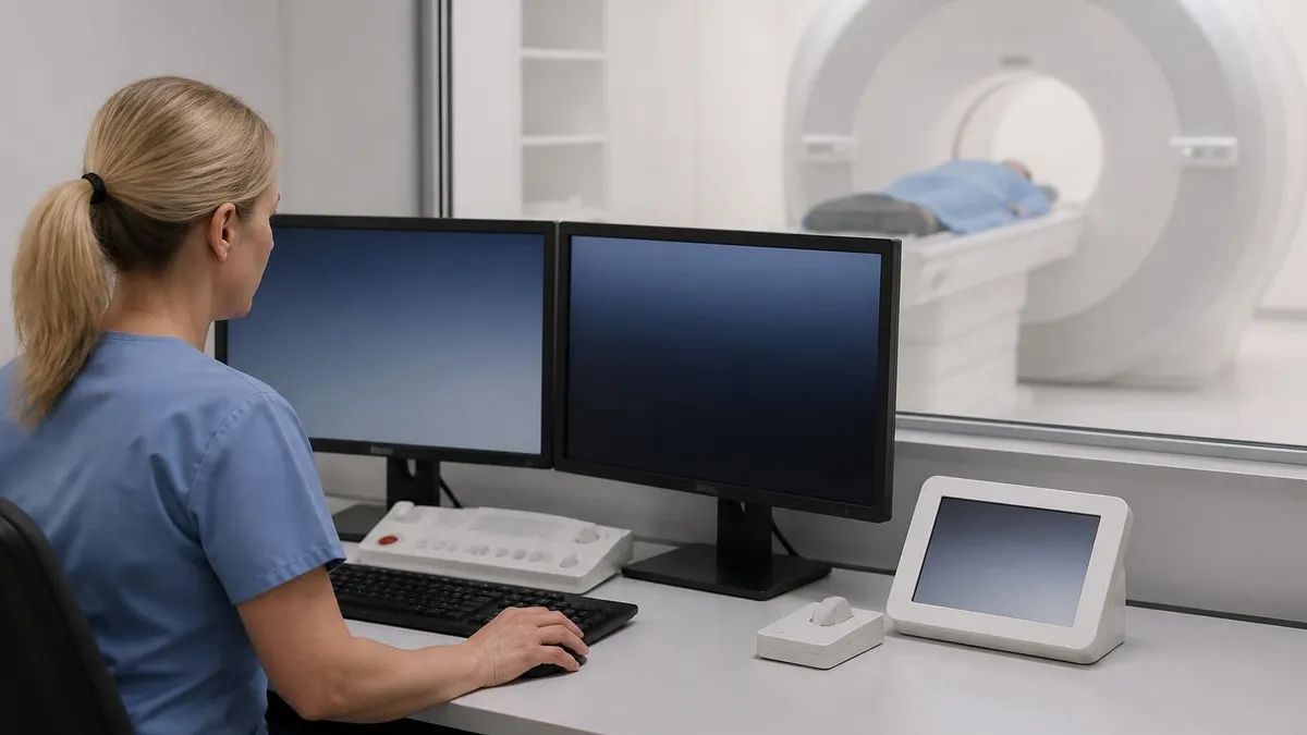
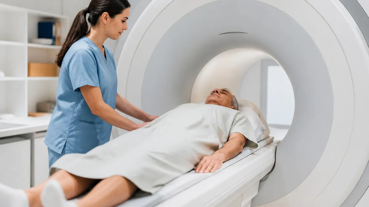
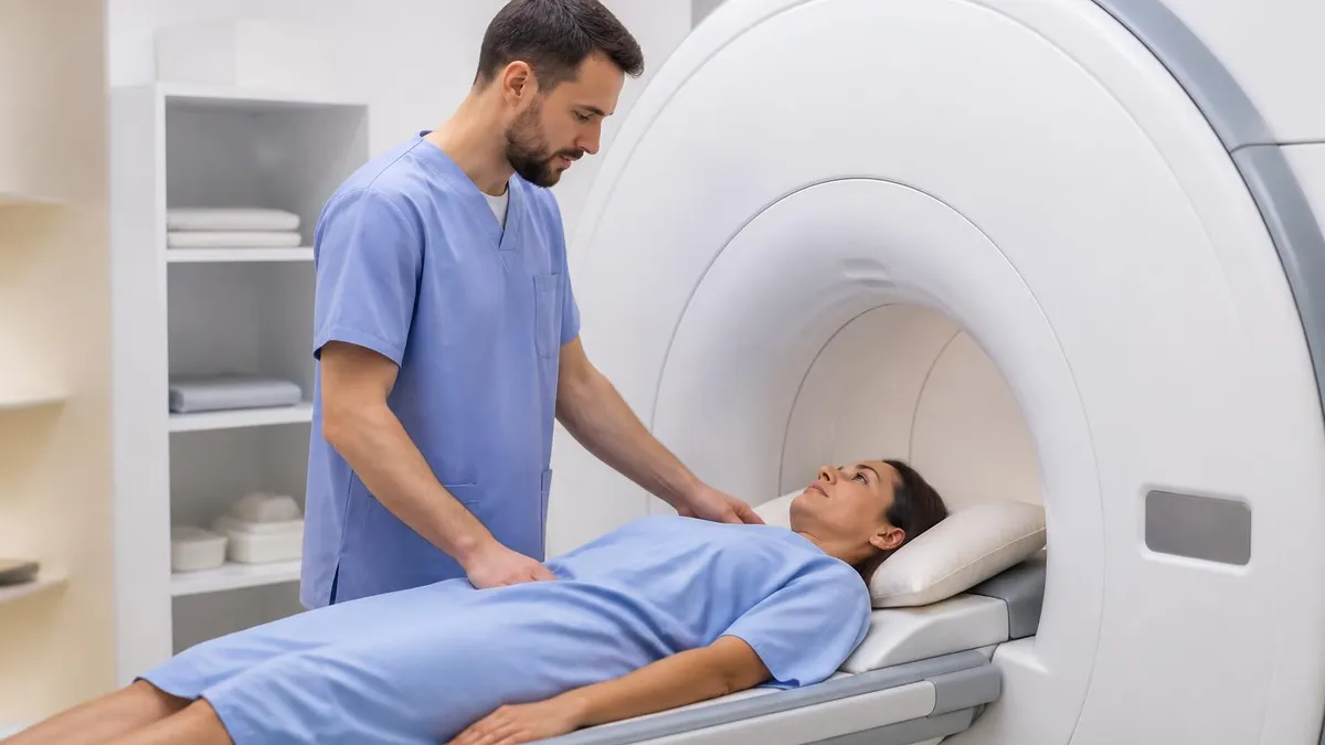
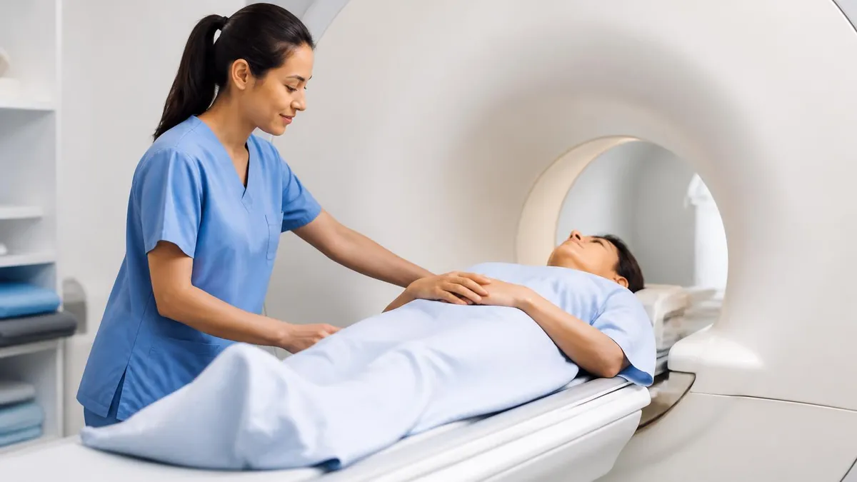
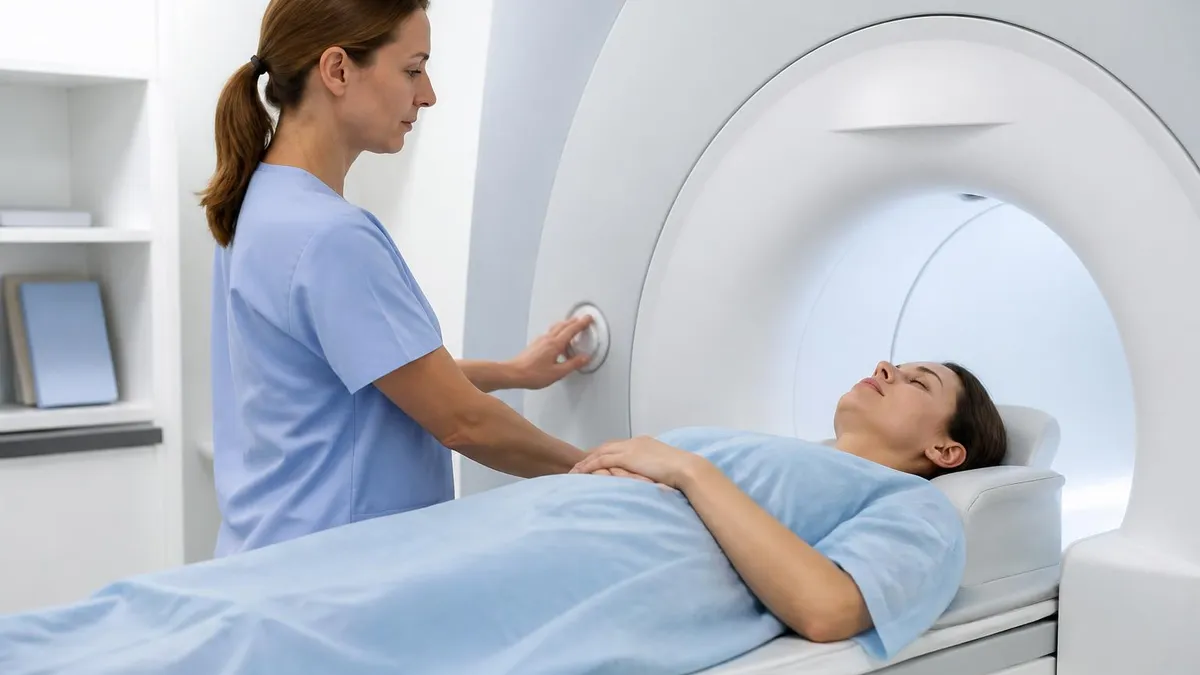
The standard shoulder MRI protocol for SLAP evaluation begins with patient positioning. The arm is placed in slight external rotation with the thumb pointing up, which separates the biceps tendon from the rotator cuff and reduces magic angle artifact at the biceps anchor. A dedicated shoulder coil is essential because body coils produce signal-to-noise ratios too low to resolve the thin fibrocartilaginous labrum. The patient is positioned supine with the affected shoulder as close to isocenter as the bore allows, accepting some asymmetric positioning to maximize image quality.
Sequence selection drives diagnostic accuracy. A typical non-contrast SLAP protocol includes axial proton density with fat saturation, coronal oblique T1 and T2 fat-saturated, sagittal oblique T2 fat-saturated, and a coronal oblique intermediate-weighted sequence. The coronal oblique plane is angled along the supraspinatus muscle belly to display the labrum in its true cross-section. Slice thickness should not exceed 3 mm, with an in-plane resolution of 0.4 mm or better to resolve the sublabral recess from a true tear.
MR arthrography elevates the diagnostic confidence substantially. Under fluoroscopic guidance, the radiologist injects 12 to 15 mL of dilute gadolinium (typically 1:200 with saline and iodinated contrast) into the glenohumeral joint. The patient is then transferred to the MRI scanner within 30 minutes before contrast diffuses into surrounding tissues. T1-weighted fat-saturated sequences in three planes demonstrate contrast tracking into any labral defect, which appears as a bright linear signal interrupting the dark labral triangle.
The ABER (abduction external rotation) position deserves special attention. By abducting the arm 90 degrees and externally rotating the forearm overhead, the inferior glenohumeral ligament tightens and pulls on the anteroinferior labrum, opening subtle Bankart-type tears that would otherwise be missed. While ABER is most useful for instability evaluation, it can also reveal anterior extension of a SLAP lesion. Most musculoskeletal centers reserve ABER imaging for arthrographic studies because it requires additional scan time.
Understanding the principles behind these protocols is easier when you review how does an MRI work and the physics of fluid-sensitive sequences. T2-weighted images use long TR and long TE values to maximize the signal difference between water-rich pathology and surrounding fat or fibrocartilage. Fat saturation chemically suppresses the bright marrow fat that would otherwise dominate the image, allowing subtle bone marrow edema adjacent to a labral tear to become visible as a hyperintense focus on the glenoid rim.
Field strength has become an active topic in SLAP imaging. 3T scanners provide approximately double the signal-to-noise ratio of 1.5T systems at equivalent scan times, allowing thinner slices and higher matrix sizes. Several published series have shown that 3T non-contrast shoulder MRI approaches the sensitivity of 1.5T arthrography for type II SLAP detection. However, 3T also amplifies susceptibility artifact near surgical hardware and metallic debris, which is why post-operative shoulders are often imaged at 1.5T using metal artifact reduction sequences.
Pulse sequence parameters must be tuned to your specific scanner. Typical coronal oblique T2 fat-saturated parameters on a 3T system include TR 4000 ms, TE 60 ms, slice thickness 3 mm, field of view 14 cm, matrix 384 by 256, and two signal averages. The technologist should always check that the saturation band is correctly positioned and that there is no aliasing wrap from the contralateral shoulder. A well-prescribed protocol produces images where the labrum, biceps anchor, and articular cartilage are all distinguishable as separate anatomical structures.
MRI Practice Test Questions
Prepare for the MRI - Magnetic Resonance Imaging exam with our free practice test modules. Each quiz covers key topics to help you pass on your first try.
MRI Knowledge
MRI Exam Questions covering Knowledge. Master MRI Test concepts for certification prep.
MRI Physics
Free MRI Practice Test featuring Physics. Improve your MRI Exam score with mock test prep.
MRI Anatomy and Pathology
MRI Test Prep for MRI Anatomy and Pathology. Practice MRI Quiz questions and boost your score.
MRI Anatomy and Positioning
MRI Questions and Answers on MRI Anatomy and Positioning. Free MRI practice for exam readiness.
MRI Contrast Agents
Free MRI Quiz on MRI Contrast Agents. MRI Exam prep questions with detailed explanations.
MRI Patient Care and Positioning
MRI Practice Questions for MRI Patient Care and Positioning. Build confidence for your MRI certification exam.
Direct vs Indirect MR Arthrography for SLAP Tear MRI
Direct MR arthrography involves fluoroscopic or ultrasound-guided injection of approximately 12 to 15 mL of dilute gadolinium contrast (1:200 with saline) directly into the glenohumeral joint capsule before MRI scanning. This technique distends the joint, separates the labral structures from surrounding tissues, and fills any communicating tear with bright signal on T1-weighted fat-saturated sequences. It remains the gold standard for SLAP detection.
Sensitivity for type II SLAP tears with direct arthrography reaches 89 to 92 percent in published series, with specificity approaching 91 percent. The technique requires fluoroscopy time, sterile injection technique, and patient cooperation. Mild post-injection discomfort is common but resolves within 24 hours. Contraindications include skin infection over the injection site, severe coagulopathy, and known gadolinium hypersensitivity.
MR Arthrography for SLAP Tear MRI: Pros and Cons
- +Highest sensitivity (90%+) for detecting type II SLAP tears
- +Joint distension separates labral fragments from glenoid rim
- +Clearly demonstrates communication between joint and tear cleft
- +Allows simultaneous diagnostic and therapeutic anesthetic injection
- +Improves detection of partial-thickness articular-side cuff tears
- +Enables ABER positioning for anterior labral evaluation
- +Provides definitive answer prior to arthroscopy
- −Requires invasive intra-articular injection
- −Adds approximately 30 minutes to total appointment time
- −Mild post-injection pain in roughly 30% of patients
- −Small risk of septic arthritis (less than 1 in 10,000)
- −Contraindicated in patients with gadolinium hypersensitivity
- −Requires fluoroscopy or ultrasound guidance equipment
SLAP Tear MRI Pre-Scan Checklist
- ✓Confirm patient has no MRI-incompatible implants or pacemaker
- ✓Screen for renal function if MR arthrography contrast is planned
- ✓Verify gadolinium allergy history and prior reactions to contrast
- ✓Position arm in neutral or slight external rotation with thumb up
- ✓Place dedicated shoulder coil snug against the shoulder joint
- ✓Center the scanner laser on the coracoid process landmark
- ✓Prescribe coronal oblique plane along the supraspinatus muscle belly
- ✓Set slice thickness to 3 mm or less with no interslice gap
- ✓Apply fat saturation to all fluid-sensitive sequences
- ✓Add ABER positioning for arthrographic studies when clinically indicated
- ✓Confirm immobilization to prevent motion during long acquisitions
- ✓Document arm rotation angle in technologist notes for the radiologist
Arm position changes labral appearance
Internal rotation of the arm during shoulder MRI can compress the superior labrum against the glenoid and mask a small type II SLAP tear. Always document the arm position. Slight external rotation with the thumb pointing up is the standard, and any deviation should be noted on the technologist worksheet so the radiologist can account for it during interpretation.
Reading a slap tear mri systematically prevents diagnostic errors. Begin every shoulder study with the axial sequence to orient yourself to the anterior, posterior, and inferior labrum. The labrum should appear as a uniformly dark triangular wedge attached to the glenoid bone. Any bright signal interrupting this triangle on a T2 or fat-saturated sequence warrants careful evaluation. The clock face system, where 12 o'clock is superior, 3 o'clock is anterior (in a right shoulder), 6 o'clock is inferior, and 9 o'clock is posterior, provides standardized terminology for tear location.
On the coronal oblique sequence, focus first on the biceps anchor at 12 o'clock. Identify the dark triangular labrum, the bright articular cartilage of the humeral head and glenoid, and the long head of the biceps tendon entering the joint. A type II SLAP appears as a hyperintense linear cleft running medially along the superior glenoid rim, extending beyond the normal sublabral recess. The tear typically extends laterally toward the biceps anchor and may show fluid signal extending into the substance of the biceps tendon itself.
Sagittal oblique images provide the bird's-eye view of the glenoid clock face. Scrolling from lateral (humeral head) to medial (glenoid), the labrum becomes visible as a thin dark ring surrounding the brighter glenoid bone. SLAP tears appear at the 10 to 2 o'clock position. Combined SLAP-Bankart lesions show additional disruption at the 3 to 5 o'clock anteroinferior labrum. Posterior extension into the 9 o'clock region indicates a more complex injury pattern, often seen in throwing athletes.
Paralabral cysts are an important secondary sign. These fluid-filled outpouchings communicate with a labral tear through a small defect and accumulate in the spinoglenoid notch or suprascapular notch. A cyst in these locations should always prompt a careful search for an underlying SLAP or posterior labral tear, even if the tear itself is not immediately apparent. Cysts can compress the suprascapular nerve and produce denervation atrophy of the infraspinatus muscle, which appears as fatty infiltration on T1 sagittal images.
The biceps anchor itself deserves dedicated evaluation. A normal biceps anchor demonstrates a continuous dark band of tendon merging with the superior labrum. In type II SLAP lesions, this dark band lifts off the glenoid with bright fluid or contrast tracking underneath. Magic angle artifact, where collagen fibers oriented at 55 degrees to the main magnetic field produce artificial hyperintensity, can mimic a tear at the biceps anchor on short TE sequences. Always confirm suspected tears on T2 long TE images where magic angle effects disappear.
The decision to recommend MR arthrography versus direct surgical evaluation depends on clinical context. Understanding mri with or without contrast helps clinicians appreciate why intra-articular gadolinium dramatically improves labral evaluation while intravenous contrast does not. Direct arthrography distends the capsule, opens labral clefts, and fills tear defects with bright signal. Intravenous gadolinium, used for tumor and inflammation evaluation, does not enter the joint capsule in sufficient concentration to outline labral structures.
Reporting a SLAP tear should include the tear type per the Snyder classification, the extent of biceps anchor involvement, the presence of any paralabral cysts, the location of the tear on the clock face, any associated rotator cuff pathology, and the presence of glenohumeral arthritis or chondral defects. Standardized reporting templates improve communication with orthopedic surgeons and help them plan arthroscopic repair versus biceps tenodesis. Mentioning the imaging confidence level (definite, probable, possible) acknowledges the limitations of imaging and guides surgical decision-making.
The sublabral foramen, sublabral recess, and Buford complex are normal anatomical variants that can be mistaken for SLAP tears. A sublabral foramen at the 2 o'clock position is present in roughly 12% of patients. The Buford complex (absent anterosuperior labrum with thickened middle glenohumeral ligament) occurs in 1.5% of patients. Misidentifying these as tears leads to unnecessary surgery.
Anatomical variants are the single most common cause of false-positive SLAP diagnoses. The sublabral recess is a normal cleft between the superior labrum and glenoid that occurs at the biceps insertion. It appears as a small, smooth-margined hyperintense line running parallel to the glenoid articular surface, typically less than 3 mm in depth. A true SLAP tear, by contrast, extends laterally beyond the biceps anchor, runs in irregular directions, and often shows displaced labral fragments or paralabral cysts. Distinguishing these two findings requires careful attention to morphology, not just signal intensity.
The Buford complex is a more dramatic variant in which the anterosuperior labrum is completely absent and replaced by a thickened, cord-like middle glenohumeral ligament. On axial images, this appears as a focal labral defect at the 1 to 3 o'clock position. The key to recognizing the variant is the cord-like middle glenohumeral ligament, which appears as a thick dark band crossing the joint. Surgical repair of a Buford complex creates iatrogenic limitation of external rotation and should never be performed.
Sublabral foramen, the third common variant, is a focal detachment of the anterosuperior labrum without disruption of the middle glenohumeral ligament. Unlike a SLAP tear, the foramen has smooth margins, is limited to the anterosuperior quadrant, and does not extend into the biceps anchor. Comparing the patient's findings to anatomical atlases and confirming the location relative to the biceps insertion prevents misidentification. Communication between the radiologist and referring surgeon is invaluable when uncertainty exists.
Magic angle phenomenon affects short TE sequences (proton density and T1) when collagen fibers in the biceps tendon or labrum are oriented at approximately 55 degrees to the main magnetic field. The resulting artificial hyperintensity can mimic a tear at the biceps anchor. The diagnostic key is to look at the T2 long TE image. True tears remain hyperintense on T2; magic angle artifact does not. Always cross-reference findings between sequences before reporting a tear at the biceps anchor.
Motion artifact is another frequent source of error. Patients with severe shoulder pain may struggle to remain still during 30 minutes of scanning. Motion creates ghosting in the phase-encoding direction, blurs labral margins, and can simulate or hide tears. Repositioning the patient, applying additional padding for comfort, and shortening sequences when possible all help reduce motion. If motion persists, the technologist should note this on the worksheet so the radiologist interprets ambiguous findings cautiously.
Hardware artifact in post-operative shoulders deserves a dedicated approach. Bioabsorbable suture anchors produce minimal artifact and allow excellent post-repair imaging. Metallic anchors, by contrast, produce signal voids that obscure the adjacent labrum. MARS protocols (Metal Artifact Reduction Sequences) use higher receiver bandwidth, view-angle tilting, and other techniques to minimize this artifact. Reviewing mri protocols specifically designed for metal-containing shoulders helps technologists deliver useful post-operative studies despite the inherent challenges.
Finally, consider the patient's clinical presentation alongside the imaging. A 55-year-old patient with insidious posterior shoulder pain and a finding of mild superior labral fraying likely has a type I degenerative SLAP that does not require repair. A 22-year-old college baseball pitcher with a frank type II detachment and a paralabral cyst is a clear surgical candidate. Imaging never exists in isolation. Always correlate the findings with the referring physician's clinical question, the patient's age and activity level, and the response to conservative therapy.
For working technologists, mastering the SLAP tear MRI workflow begins with consistent positioning. Develop a routine: thumb up, arm slightly externally rotated, padding under the elbow for comfort, dedicated shoulder coil tightly placed, laser centered at the coracoid. Repeating this routine on every shoulder study builds muscle memory and reduces variability between technologists. Variability is the enemy of accurate diagnosis, because a borderline tear that appears clearly on one scan may be invisible on the next if positioning differs.
For registry candidates preparing for the ARRT MRI examination, focus on understanding the physics behind fluid-sensitive sequences. Know that T2-weighted sequences use long TR and long TE to maximize signal from fluid-containing pathology. Understand how fat saturation works through chemical shift selective pulses or short tau inversion recovery (STIR). Recognize that MR arthrography uses T1-weighted sequences because dilute gadolinium shortens T1 relaxation and appears bright on T1, not T2.
For radiology residents, develop a systematic reading approach. Always evaluate the labrum on at least two planes before calling a tear. Confirm signal abnormality on both proton density and T2 fat-saturated sequences to exclude magic angle artifact. Measure the depth and extent of any labral cleft. Compare to the contralateral shoulder when available. Document findings using standardized terminology and the Snyder classification system. Always include differential diagnoses for ambiguous findings.
For orthopedic surgeons interpreting reports, recognize that radiologists communicate diagnostic confidence through phrases like "definite type II SLAP," "probable type II SLAP," or "findings concerning for SLAP but anatomical variant cannot be excluded." These distinctions matter for surgical planning. A definite tear in a young athlete with mechanical symptoms warrants arthroscopic evaluation. An equivocal finding in an older patient with global pain may warrant conservative management before surgical exploration.
Understanding the historical evolution of magnetic resonance imaging illuminates why current protocols exist. The history of MRI shows how labral imaging evolved from low-field 0.3T systems with poor labral resolution to modern 3T scanners with sub-millimeter detail. Each technological advance, from dedicated extremity coils to parallel imaging acceleration, has improved SLAP detection sensitivity and shortened scan times. Knowing this history helps current practitioners appreciate why protocol details matter.
Patient communication is often overlooked but essential. Many patients arriving for shoulder MRI are anxious about the noisy bore environment, the long scan time, and the potential for arthrographic injection. A brief pre-scan conversation explaining what to expect, what positions will be required, and how long the scan will take dramatically improves cooperation and image quality. Patients who understand they need to remain still for specific sequences are far more compliant than those facing unexpected delays.
Finally, remember that imaging findings must always be correlated with clinical examination. The O'Brien active compression test, the crank test, and the biceps load tests all have specific patterns of pain that suggest SLAP pathology. A positive imaging finding in a patient with positive clinical tests is far more meaningful than an isolated imaging finding. Imaging confirms clinical suspicion; it should not generate diagnoses in isolation. This is why orthopedic surgeons value technologists and radiologists who provide accurate, well-documented studies that support clinical decision-making.
MRI Questions and Answers
MRI MARS Protocol: Complete Guide to Metal Artifact Reduction Sequences for Orthopedic Imaging
How Does an MRI Work: Magnetic Resonance Imaging Explained
MRI With or Without Contrast: What to Expect
The History of MRI: From Discovery to Modern Medicine
What's the Difference Between MRI and CT Scan? A Complete Comparison Guide
About the Author
Medical Laboratory Scientist & Clinical Certification Expert
Johns Hopkins UniversityDr. Sandra Kim holds a PhD in Clinical Laboratory Science from Johns Hopkins University and is certified as a Medical Technologist (MT) and Medical Laboratory Scientist (MLS) through ASCP. With 16 years of clinical laboratory experience spanning hematology, microbiology, and molecular diagnostics, she prepares candidates for ASCP board exams, MLT, MLS, and specialist certification tests.
Join the Discussion
Connect with other students preparing for this exam. Share tips, ask questions, and get advice from people who have been there.
View discussion (6 replies)