PET MRI: Complete Guide to Hybrid PET/MRI Imaging Technology 2026 July
PET MRI combines molecular imaging with high-resolution MRI. ✍🏼 Learn how hybrid PET/MRI works, clinical uses, costs, and what to expect during your scan.
PET MRI is a hybrid imaging modality that fuses positron emission tomography with magnetic resonance imaging in a single examination, giving clinicians both metabolic and anatomical information simultaneously. Unlike traditional PET/CT, a PET MRI system captures soft-tissue detail with exquisite contrast while delivering the molecular sensitivity that nuclear medicine has long provided. The technology emerged commercially around 2010 and has since become a cornerstone for oncology, neurology, cardiology, and pediatric imaging, particularly when minimizing ionizing radiation matters.
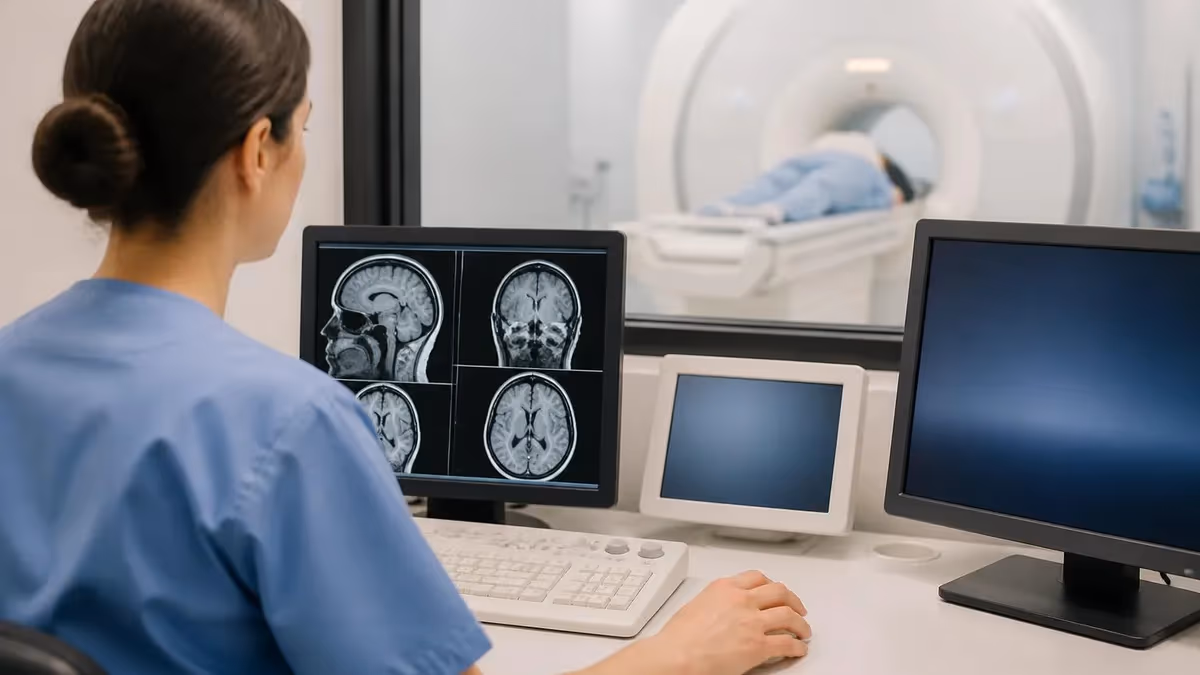
The appeal of PET MRI lies in the way two complementary signals are acquired and reconstructed together. PET shows where injected radiotracers like fluorodeoxyglucose accumulate, highlighting metabolically active tissue such as tumors, inflammation, or epileptic foci. MRI delivers detailed anatomy, perfusion maps, diffusion characteristics, and functional brain activity. When the two datasets are co-registered in the same gantry, motion mismatch shrinks dramatically and lesion localization becomes far more reliable than sequentially acquired studies.
Adoption of hybrid PET MRI has been steady but selective because the scanners are expensive, technically complex, and require specialized staff. Academic medical centers and large cancer hospitals were the earliest adopters, followed by neuroscience research institutes and pediatric programs. Today, manufacturers including Siemens, GE HealthCare, and United Imaging continue to refine integrated platforms with improved attenuation correction, time-of-flight detectors, and silicon photomultiplier technology that boost image quality and shorten scan times.
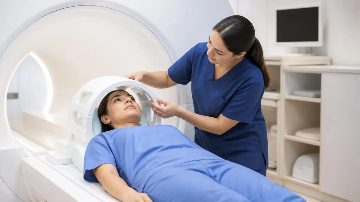
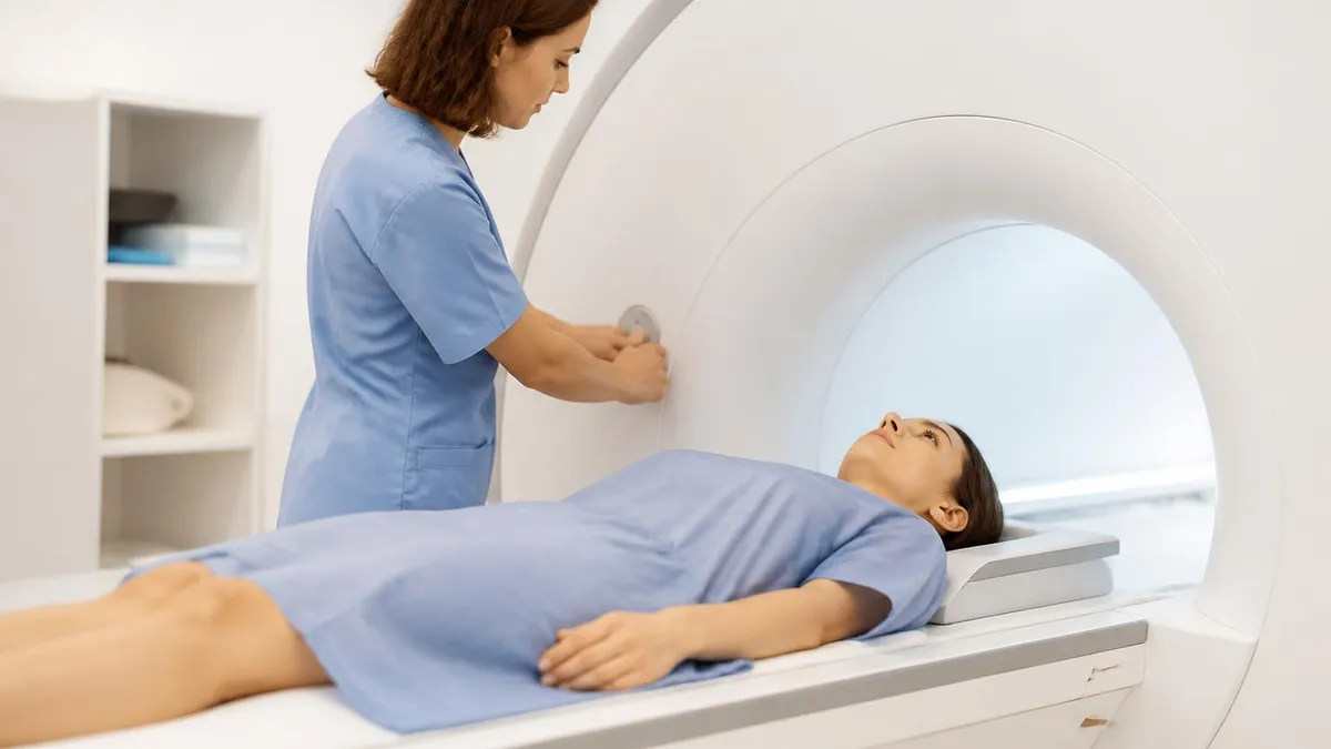
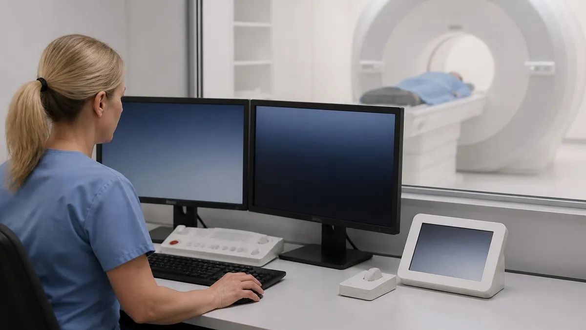
From a patient's perspective, a PET MRI visit feels similar to a traditional MRI but with the addition of a small intravenous radiotracer injection roughly an hour before scanning. The combined exam typically lasts 45 to 90 minutes depending on the protocol, body region, and clinical question. Patients lie still inside the magnet bore while sequences are acquired, and contrast agents may be administered if needed for specific MRI pulse sequences. Most patients tolerate the exam well, though claustrophobia and breath-holding requirements remain consistent challenges.
Clinical evidence supporting PET MRI continues to expand across multiple specialties. In oncology, the modality excels at staging prostate cancer, characterizing liver lesions, evaluating head and neck tumors, and detecting bone marrow involvement. In neurology, it has transformed presurgical epilepsy mapping, dementia workup, and brain tumor characterization. Cardiology applications include myocardial viability assessment and sarcoidosis evaluation. Pediatric oncology programs particularly value PET MRI because it can reduce cumulative radiation exposure compared with repeated PET/CT studies.
Despite its strengths, PET MRI is not a universal replacement for PET/CT. CT remains superior for lung nodule detection, calcified lesions, and rapid whole-body coverage. Workflow throughput on PET MRI is slower, reimbursement varies, and not every radiology department has the staffing depth to support hybrid interpretation. Still, when the clinical question calls for high soft-tissue contrast paired with molecular sensitivity, PET MRI is often the most accurate diagnostic tool available, and its role continues to grow as protocols mature and tracer libraries expand.
This guide explores how PET MRI works, who benefits most, the technical underpinnings of the scanner, what to expect before and after the exam, and how it compares to other modalities. Whether you are a technologist preparing for the registry, a referring physician evaluating the modality for a patient, or a curious learner researching how does an MRI work alongside nuclear medicine, you will find practical, evidence-based answers throughout the sections that follow.
PET MRI by the Numbers
How PET MRI Technology Works
Modern PET MRI scanners use silicon photomultipliers or avalanche photodiodes that function inside strong magnetic fields, unlike older photomultiplier tubes. The detectors sit within the MRI gantry, allowing truly simultaneous acquisition rather than sequential scans on separate machines.
Because MRI cannot directly measure photon attenuation like CT, PET MRI uses MR-based attenuation correction maps derived from Dixon sequences or ultrashort echo time imaging. These maps segment tissues into air, lung, fat, and soft tissue, then assign attenuation values to correct PET data.
Time-of-flight PET measures the difference in arrival times of paired annihilation photons to localize events more precisely along each line of response. This boosts signal-to-noise ratio significantly, particularly in larger patients, and shortens acquisition times without sacrificing diagnostic quality.
Unlike PET/CT systems that scan sequentially, integrated PET MRI captures both datasets at the same time and in the same anatomical position. This eliminates registration errors caused by patient motion, bladder filling, or breathing differences between exams, improving lesion localization accuracy.
PET MRI combines metabolic information from radiotracers with multiple MRI contrasts including T1, T2, diffusion-weighted imaging, dynamic contrast enhancement, and spectroscopy. This multi-parametric approach provides a richer dataset for characterizing tumors, inflammatory lesions, and neurological diseases in a single visit.
Clinical applications for PET MRI span oncology, neurology, cardiology, musculoskeletal imaging, and pediatrics, with each specialty leveraging the modality's strengths differently. In oncology, the most common indications include prostate cancer staging with PSMA tracers, hepatocellular carcinoma evaluation, head and neck cancer staging, gynecologic malignancies, and pediatric solid tumors. The high soft-tissue contrast of MRI helps distinguish tumor from adjacent normal tissue, while PET highlights metabolically active disease that may be missed on anatomical imaging alone.
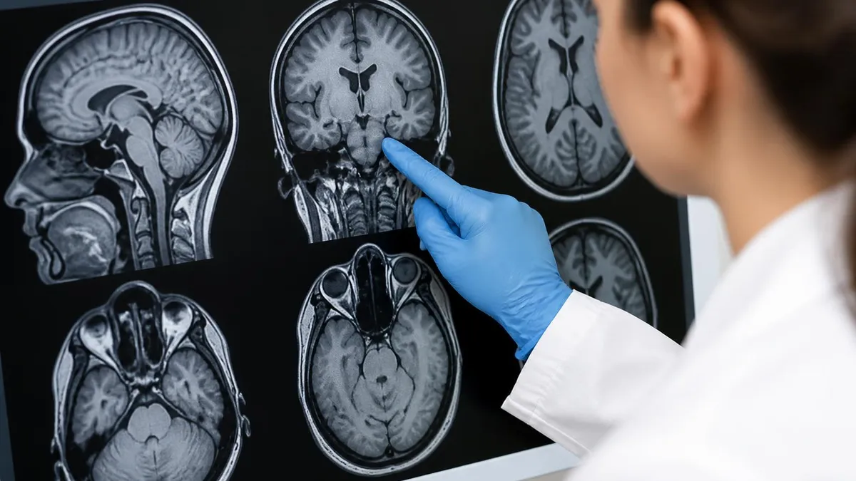
Neurological applications have transformed how clinicians approach complex brain disorders. Presurgical epilepsy evaluation benefits enormously from simultaneous PET hypometabolism mapping fused with high-resolution structural MRI, allowing surgeons to localize seizure foci that would be invisible on either modality alone. Dementia workup uses amyloid and tau PET tracers alongside volumetric MRI to differentiate Alzheimer's disease from frontotemporal dementia, vascular cognitive impairment, and Lewy body disease. Brain tumor recurrence versus radiation necrosis is another classic indication.
Cardiac PET MRI offers unique value for evaluating myocardial viability, cardiac sarcoidosis, amyloidosis, and inflammatory myocarditis. The combination of late gadolinium enhancement, T1 and T2 mapping, and FDG metabolism provides comprehensive tissue characterization that no single modality can match. Patients with suspected sarcoidosis particularly benefit because PET MRI can simultaneously assess active inflammation, fibrosis burden, and cardiac function in one sitting, guiding immunosuppressive therapy decisions with confidence.
Musculoskeletal PET MRI is gaining traction for evaluating bone marrow involvement in lymphoma and myeloma, prosthetic joint infection, osteomyelitis, and primary bone tumors. Whole-body diffusion-weighted MRI combined with FDG PET provides sensitive detection of marrow disease without the radiation burden of repeated CT or skeletal surveys. For complex orthopedic infections, the modality helps distinguish active inflammation from chronic post-surgical changes when other imaging is equivocal or contradictory.
Pediatric oncology programs have embraced PET MRI as a radiation-sparing alternative to PET/CT for children with lymphoma, sarcoma, neuroblastoma, and other solid tumors. Because pediatric patients often require multiple staging and follow-up studies over months or years, cumulative radiation exposure becomes a real concern. PET MRI reduces dose by 50 to 80 percent compared with PET/CT while delivering equivalent or superior diagnostic accuracy, making it increasingly the preferred staging modality at major children's hospitals.
Inflammatory and infectious diseases represent another expanding indication. Large vessel vasculitis, inflammatory bowel disease, fever of unknown origin, and complex infections all benefit from the molecular sensitivity of FDG PET combined with anatomical detail from MRI. Rheumatologists increasingly request PET MRI for difficult cases where laboratory and clinical findings remain ambiguous, and the modality has helped guide biopsy targeting and treatment monitoring in challenging autoimmune presentations across multiple organ systems.
Despite expanding indications, appropriate use criteria still guide PET MRI ordering. Insurance pre-authorization, scanner availability, and clinical expertise vary widely across regions. Many centers reserve PET MRI for cases where PET/CT or MRI alone would be inadequate, or where reducing radiation exposure justifies the added cost and time. Understanding whats difference between mri and ct scan is essential for clinicians deciding which hybrid platform best answers a given clinical question.
MRI Practice Test Questions
Prepare for the MRI - Magnetic Resonance Imaging exam with our free practice test modules. Each quiz covers key topics to help you pass on your first try.
MRI Knowledge
MRI Exam Questions covering Knowledge. Master MRI Test concepts for certification prep.
MRI Physics
Free MRI Practice Test featuring Physics. Improve your MRI Exam score with mock test prep.
MRI Anatomy and Pathology
MRI Test Prep for MRI Anatomy and Pathology. Practice MRI Quiz questions and boost your score.
MRI Anatomy and Positioning
MRI Questions and Answers on MRI Anatomy and Positioning. Free MRI practice for exam readiness.
MRI Contrast Agents
Free MRI Quiz on MRI Contrast Agents. MRI Exam prep questions with detailed explanations.
MRI Patient Care and Positioning
MRI Practice Questions for MRI Patient Care and Positioning. Build confidence for your MRI certification exam.
PET MRI vs PET CT vs MRI Alone
PET MRI delivers unmatched soft-tissue contrast for evaluating brain, liver, pelvis, and musculoskeletal disease. The simultaneous acquisition eliminates registration errors between metabolic and anatomical data, which is especially valuable for moving organs or small lesions near boundaries. Radiation exposure is significantly lower than PET/CT, making it the preferred choice for pediatric patients and young adults requiring serial imaging studies over months or years of treatment monitoring.
Functional MRI sequences add layers of information no other hybrid modality can match. Diffusion-weighted imaging quantifies cellularity, dynamic contrast-enhanced sequences map perfusion, and spectroscopy reveals metabolite concentrations. When combined with PET tracer uptake, clinicians can characterize tumors, inflammation, and treatment response with remarkable depth, often resolving diagnostic dilemmas that single-modality imaging cannot answer with sufficient confidence for clinical decisions.
Is PET MRI Right for Your Clinical Question?
- +Significantly lower radiation dose than PET/CT, ideal for pediatrics and young adults
- +Superior soft-tissue contrast for brain, liver, pelvis, and musculoskeletal imaging
- +Simultaneous acquisition eliminates motion mismatch between metabolic and anatomical data
- +Multi-parametric MRI sequences add diffusion, perfusion, and spectroscopy information
- +Excellent for presurgical epilepsy mapping and complex dementia workups
- +One appointment instead of two separate PET and MRI exams
- −Limited availability with only about 150 installed systems across the United States
- −Higher cost than PET/CT, often $3,000 to $8,000 per exam
- −Longer scan times that can be challenging for claustrophobic or unstable patients
- −MR-based attenuation correction less accurate than CT for bone and lung
- −Insurance pre-authorization can be more difficult than for PET/CT
- −Requires specialized technologists trained in both MRI and nuclear medicine workflows
PET MRI Patient Preparation Checklist
- ✓Fast for 4 to 6 hours before the exam, drinking only water unless instructed otherwise
- ✓Avoid strenuous exercise for 24 hours before the scan to reduce muscle uptake
- ✓Inform the technologist about diabetes and recent blood glucose levels
- ✓Remove all metal objects including jewelry, hearing aids, dentures, and piercings
- ✓Complete a comprehensive MRI safety screening for implants, pacemakers, and metallic foreign bodies
- ✓Notify staff of pregnancy, breastfeeding, or possible pregnancy before tracer injection
- ✓Arrive 60 to 90 minutes early to allow time for tracer injection and uptake
- ✓Stay still and relaxed during the uptake phase, avoiding talking, chewing, or movement
- ✓Empty your bladder immediately before the scan to improve pelvic image quality
- ✓Plan for the entire visit to take 2 to 3 hours from check-in to discharge
When PET MRI Changes Management
PET MRI most clearly outperforms PET/CT when the clinical question involves brain, liver, pelvis, head and neck, or pediatric imaging. For lung-predominant disease, PET/CT remains superior. Ordering the right modality the first time saves weeks and avoids redundant radiation exposure.
Radiotracers used in PET MRI are identical to those used in PET/CT and span a growing library of molecules targeting specific biological pathways. Fluorodeoxyglucose remains the most common, leveraging glucose metabolism to highlight tumors, infection, and inflammation. Newer tracers including PSMA ligands for prostate cancer, DOTATATE for neuroendocrine tumors, fluciclovine for prostate recurrence, and amyloid and tau tracers for dementia have dramatically expanded the diagnostic reach of hybrid imaging in oncology and neurology.
Tracer choice depends entirely on the clinical question and is selected jointly by the referring physician, radiologist, and nuclear medicine team. PSMA PET MRI, for example, has become a preferred staging modality for high-risk prostate cancer because it combines exquisite pelvic soft-tissue contrast from MRI with the molecular specificity of prostate-specific membrane antigen targeting. Bone metastases, lymph node involvement, and local recurrence are all visualized with sensitivity that exceeds conventional imaging by significant margins in published comparative studies.
Doses are calculated based on patient weight and tracer-specific protocols, with most adult FDG doses ranging from 5 to 15 millicuries. Pediatric dosing follows weight-based scaling and is carefully optimized to minimize radiation while maintaining diagnostic quality. The injected activity, time from injection to scan, and physiologic biodistribution all influence image interpretation, so technologists carefully document timing and any deviations from protocol that might affect quantitative analysis like standardized uptake value calculations.
Gadolinium-based MRI contrast agents are often administered during PET MRI exams to enhance specific sequences, particularly for brain, liver, and cardiac imaging. Modern macrocyclic agents like gadobutrol and gadoterate carry low risk of nephrogenic systemic fibrosis even in patients with mild to moderate kidney disease, though screening for renal function remains standard. Some protocols use hepatobiliary-specific contrast agents to characterize liver lesions, providing additional anatomical and functional information layered onto the PET data acquired simultaneously.
Patient preparation around tracer kinetics matters significantly for image quality. Fasting reduces background glucose competition for FDG uptake, allowing tumors and inflammatory lesions to stand out against suppressed normal tissue. For brown fat suppression in pediatric patients, warm blankets and ambient temperature control reduce physiologic tracer uptake that can obscure pathology. Blood glucose levels above 200 milligrams per deciliter typically require rescheduling because hyperglycemia significantly degrades FDG image quality and quantitative accuracy across all body regions.
Safety considerations combine the standard MRI checklist with nuclear medicine precautions. Patients with non-MRI-conditional pacemakers, certain cochlear implants, or retained metallic foreign bodies cannot undergo PET MRI even when PET/CT would be feasible. Pregnant patients generally avoid both ionizing radiation from tracers and gadolinium contrast unless the clinical benefit clearly outweighs theoretical risks. Breastfeeding interruption guidelines depend on the specific tracer used and follow established Society of Nuclear Medicine and Molecular Imaging recommendations.
Post-scan instructions are straightforward for most patients. Increased fluid intake helps flush residual tracer through the urinary system, and close contact with pregnant women and young children is typically minimized for several hours depending on tracer half-life. Driving, working, and normal activities resume immediately after discharge, and side effects are exceptionally rare. The injected activity decays naturally and is excreted within 24 hours for most commonly used radiotracers, leaving no lasting radioactive burden on the patient.
All PET MRI patients must complete thorough MRI safety screening because the strong magnetic field is always on. Pacemakers, implanted cardioverter defibrillators, cochlear implants, and certain aneurysm clips may be absolute contraindications. Even MRI-conditional devices require verification of model and conditions before entering the scanner room.
The cost landscape for PET MRI in the United States reflects both the capital investment and operational complexity of these hybrid systems. A new integrated scanner typically costs between $5 and $8 million to purchase and install, with annual maintenance contracts adding hundreds of thousands of dollars. Patients without insurance face self-pay charges ranging from $3,000 to $8,000 depending on the body region scanned, tracer used, and contrast administration. Insurance coverage has improved but still varies significantly by carrier, indication, and geographic region.
Reimbursement codes for PET MRI continue to evolve as the modality matures. Medicare and most commercial payers cover the modality for established oncologic indications when ordered through appropriate use criteria pathways, though pre-authorization remains common. Bundled codes covering both the PET and MRI components are gradually replacing fragmented billing, simplifying revenue cycle management for hospitals. Some indications, particularly novel tracers for prostate cancer or amyloid imaging in dementia, have specific coverage rules that referring physicians must understand to avoid surprise patient bills.
Access to PET MRI remains concentrated at academic medical centers, large cancer hospitals, and select tertiary referral sites. Approximately 150 systems are installed across the United States as of 2025, compared with thousands of PET/CT and standalone MRI scanners. Rural and community patients often travel substantial distances or settle for PET/CT when PET MRI would be ideal. Telehealth consultations and regional referral networks help bridge this gap, but geographic disparities in access remain a persistent challenge for equitable care delivery nationwide.
Workflow optimization has improved dramatically since early PET MRI installations a decade ago. Modern scanners offer continuous bed motion, shortened MR protocols designed specifically for hybrid use, and advanced reconstruction algorithms that reduce scan times. Time-of-flight detectors and digital silicon photomultipliers allow lower tracer doses and faster acquisitions without sacrificing diagnostic quality. Some centers now complete whole-body PET MRI in 30 to 45 minutes, approaching the throughput of traditional PET/CT while delivering superior soft-tissue characterization across multiple body regions.
Artificial intelligence is reshaping PET MRI interpretation and acquisition workflows. Deep learning algorithms now generate attenuation correction maps from MRI data with accuracy approaching CT-based methods, reconstruct lower-dose PET images with preserved quality, and assist radiologists in detecting and characterizing lesions. Automated structured reporting, treatment response quantification, and longitudinal tracking tools are becoming standard features. These advances reduce variability between readers and help less experienced radiologists deliver consistent, high-quality interpretations across the growing range of clinical indications.
Looking forward, the future of PET MRI is bright across multiple dimensions. Total-body PET MRI prototypes capable of imaging the entire patient in a single bed position promise dramatic reductions in scan time and tracer dose. New theranostic pairings allow imaging and treatment with related molecules, particularly in prostate and neuroendocrine cancers. Expanding tracer libraries targeting fibroblast activation protein, hypoxia, and immune cell populations will open new clinical applications. Reviewing history of mri shows how rapidly this technology has progressed, and PET MRI represents the next major evolution.
For technologists and radiologists considering specialization in PET MRI, the field offers tremendous career opportunities. Dual-credentialed staff with both MRI and nuclear medicine certifications are in high demand, with competitive salaries reflecting their specialized skill set. Academic centers actively recruit faculty radiologists with hybrid imaging expertise, and industry roles in scanner development, clinical applications, and AI software are expanding rapidly. The combination of clinical impact, technical depth, and ongoing innovation makes PET MRI one of the most exciting subspecialties in modern medical imaging today.
Preparing for a PET MRI exam, whether as a patient, technologist, or referring clinician, requires attention to several practical details that significantly impact image quality and diagnostic accuracy. Patients should follow fasting instructions precisely, arrive with comfortable clothing free of metal, and bring a list of current medications and prior imaging studies. Diabetics need special coordination because blood glucose must be controlled to acceptable levels before FDG injection, often requiring morning appointments and medication adjustments planned with the referring physician and imaging staff.
For technologists, mastering the dual workflow of MRI safety screening and radiopharmaceutical handling is essential. The MRI suite must always be approached as a Zone 4 environment where the magnet is on continuously, even during tracer injection and uptake phases. Lead shielding strategies that work in nuclear medicine departments do not translate directly to MRI environments because ferromagnetic materials cannot enter the magnet room. Specialized non-ferromagnetic infusion pumps, IV poles, and patient transport equipment are standard fixtures in modern PET MRI departments.
Quality control protocols for PET MRI systems are more complex than for either modality alone. Daily checks include verification of detector performance, attenuation correction map accuracy, coil function, and gradient stability. Weekly and monthly procedures evaluate calibration phantoms designed specifically for hybrid imaging. Documentation requirements support accreditation through bodies like the American College of Radiology and the Intersocietal Accreditation Commission, both of which have established standards for PET MRI program quality and reporting consistency across diverse clinical settings.
Image interpretation requires comfortable familiarity with both molecular and anatomical findings. Radiologists typically read PET MRI studies on dedicated workstations that display fused PET and MR images alongside individual sequences. Quantitative tools including standardized uptake values, apparent diffusion coefficients, and time-intensity curves support clinical decisions. Many programs use dual-reader workflows or joint nuclear medicine and body imaging interpretation to leverage subspecialty expertise. Structured reporting templates help ensure consistent communication of findings to referring clinicians across complex multi-system examinations.
Patient communication around PET MRI deserves thoughtful attention because the exam involves both injected radioactivity and prolonged scanner time. Clear explanations of the safety profile, expected sensations, and post-scan recommendations reduce anxiety and improve cooperation. Many programs use video tours, written instructions, and phone calls the day before to prepare patients effectively. For pediatric patients, child life specialists and detailed pre-visit education materials significantly improve the experience and reduce the need for sedation or repeat studies due to motion artifacts.
Continuing education in PET MRI is critical because the field evolves rapidly with new tracers, protocols, and software releases. Society of Nuclear Medicine and Molecular Imaging, Radiological Society of North America, and International Society for Magnetic Resonance in Medicine all offer dedicated PET MRI courses and meeting sessions. Subscription-based learning platforms, vendor training, and peer-reviewed literature complement formal continuing medical education. Staying current with appropriate use criteria, reporting standards, and emerging applications ensures programs deliver the highest quality care to patients across all indications.
Finally, building a successful PET MRI program requires strong multidisciplinary collaboration between radiology, nuclear medicine, oncology, neurology, cardiology, and pediatrics. Regular tumor boards, case conferences, and protocol review meetings keep clinical pathways aligned with the latest evidence. Investment in staff training, equipment maintenance, and IT infrastructure pays dividends in patient throughput, diagnostic accuracy, and physician satisfaction. The most successful programs treat PET MRI as a strategic clinical asset rather than just an expensive scanner, leveraging it to answer the hardest diagnostic questions in modern medicine.
MRI Questions and Answers
MRI MARS Protocol: Complete Guide to Metal Artifact Reduction Sequences for Orthopedic Imaging
How Does an MRI Work: Magnetic Resonance Imaging Explained
MRI With or Without Contrast: What to Expect
The History of MRI: From Discovery to Modern Medicine
What's the Difference Between MRI and CT Scan? A Complete Comparison Guide
About the Author
Medical Laboratory Scientist & Clinical Certification Expert
Johns Hopkins UniversityDr. Sandra Kim holds a PhD in Clinical Laboratory Science from Johns Hopkins University and is certified as a Medical Technologist (MT) and Medical Laboratory Scientist (MLS) through ASCP. With 16 years of clinical laboratory experience spanning hematology, microbiology, and molecular diagnostics, she prepares candidates for ASCP board exams, MLT, MLS, and specialist certification tests.
Join the Discussion
Connect with other students preparing for this exam. Share tips, ask questions, and get advice from people who have been there.
View discussion (6 replies)