MRI That Freezes Tumors: How Cryoablation Imaging Guides Precision Cancer Treatment
MRI that freezes tumors uses real-time imaging to guide cryoablation, freezing cancer cells with sub-millimeter precision. Learn how the procedure works. 🗨️
The phrase mri that freezes tumors refers to MRI-guided cryoablation, a minimally invasive cancer treatment that uses real-time magnetic resonance imaging to direct ultra-thin probes into tumors and freeze them at temperatures approaching minus 150 degrees Celsius. Unlike traditional surgery, this procedure leaves the surrounding tissue largely intact, relies on imaging rather than scalpels, and can often be completed in a single outpatient session lasting two to four hours from start to finish.
The technology emerged from a convergence of two mature fields: cryosurgery, which dates to the 1960s, and high-field MRI, which became clinically dominant in the 1990s. What changed in the last decade is the development of MRI-compatible cryoprobes made from titanium and zirconium alloys that do not distort the magnetic field. Radiologists can now place needles, monitor the growing ice ball in real time, and confirm complete tumor coverage without ever removing the patient from the bore.
This procedure is particularly valuable for patients who are poor surgical candidates due to age, comorbidities, or tumor location near critical structures. Kidney tumors smaller than four centimeters, certain liver metastases, painful bone lesions, low-risk prostate cancers, and select breast fibroadenomas are now routinely treated this way at major academic centers. The American College of Radiology has issued appropriateness criteria endorsing MRI-guided cryoablation for several of these indications.
The clinical appeal goes beyond avoiding incisions. Because MRI shows the ice ball as a clearly demarcated signal void with a millimeter-precise margin, the interventionalist can verify in real time that the freeze zone extends at least five millimeters beyond the tumor edge. That margin is the difference between a durable cure and local recurrence. CT-guided cryoablation, by contrast, must infer ice ball borders from indirect density changes, which is less reliable in soft tissue. Reviewing common MRI findings helps technologists understand what radiologists look for during these guided procedures.
Patients typically experience mild discomfort rather than acute pain, since freezing nerve endings produces a brief sting followed by numbness. Recovery times are measured in days rather than weeks, and most people return to normal activity within forty-eight to seventy-two hours. Hospital stays, when needed at all, are usually a single overnight observation rather than the multi-day admission required after open surgical resection.
For MRI technologists, understanding this procedure is increasingly part of the job. Interventional MRI suites are no longer rare, and registry exams from ARRT and ARMRIT now include questions about real-time imaging sequences, probe artifact recognition, and the safety considerations specific to cryoablation. This article explains the procedure end to end, from candidate selection through follow-up imaging, and equips you with the practical knowledge you need to assist effectively or to answer board-style questions confidently.
Whether you are a technologist preparing for advanced credentialing, a patient researching treatment options, or a student studying interventional radiology, the next sections will walk you through the physics, the workflow, the indications, and the evidence base behind one of the most elegant applications of MRI in modern medicine.
MRI-Guided Cryoablation by the Numbers
The Procedure Workflow Step by Step
Pre-Procedure Planning MRI
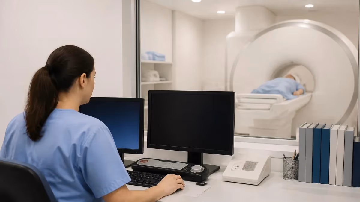
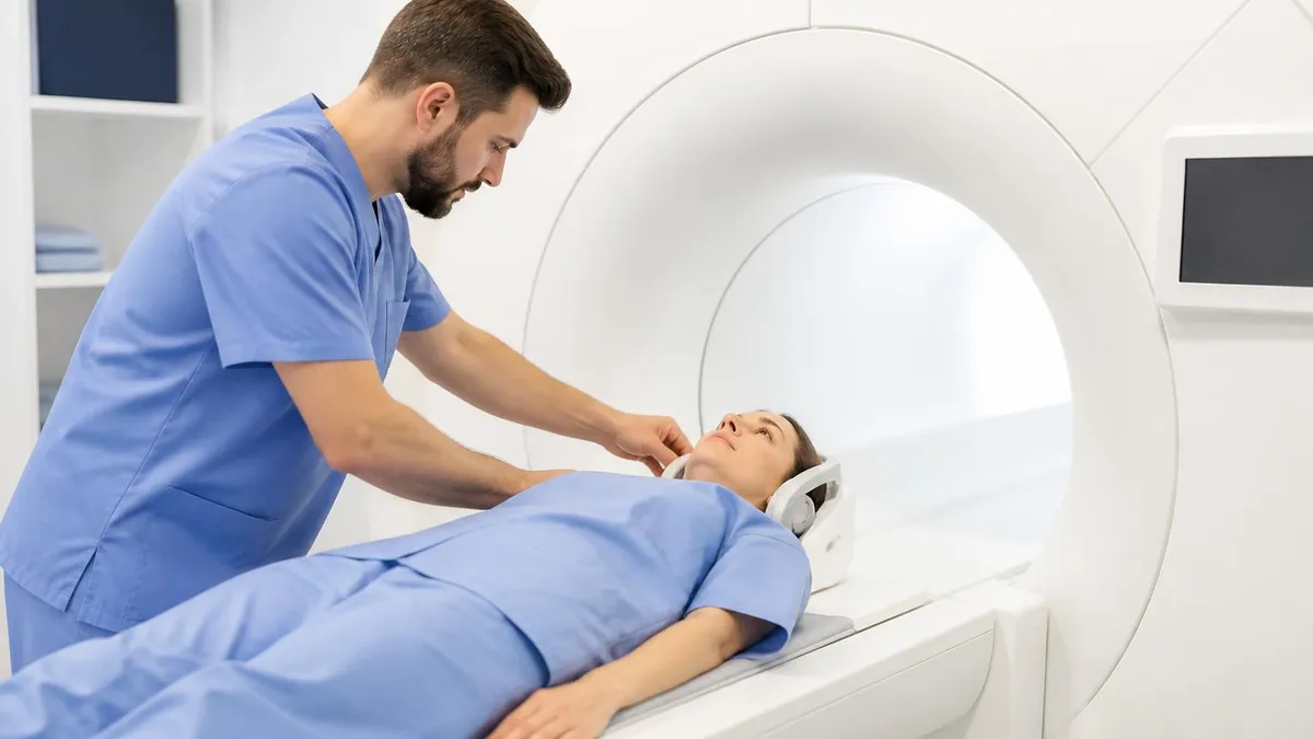
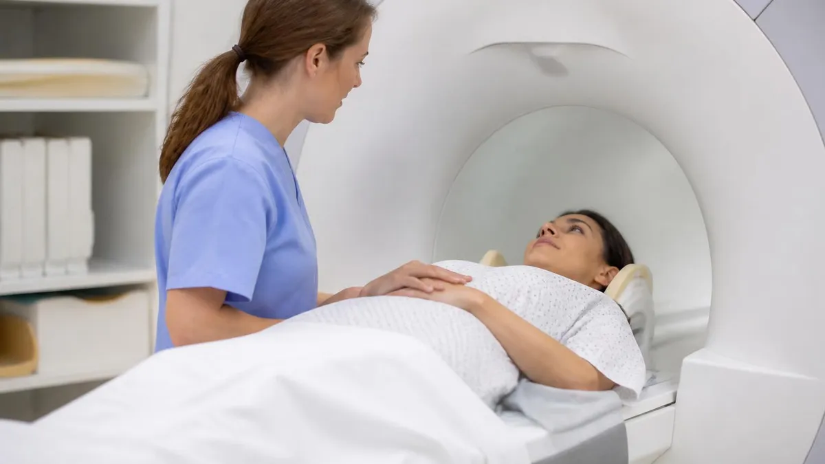
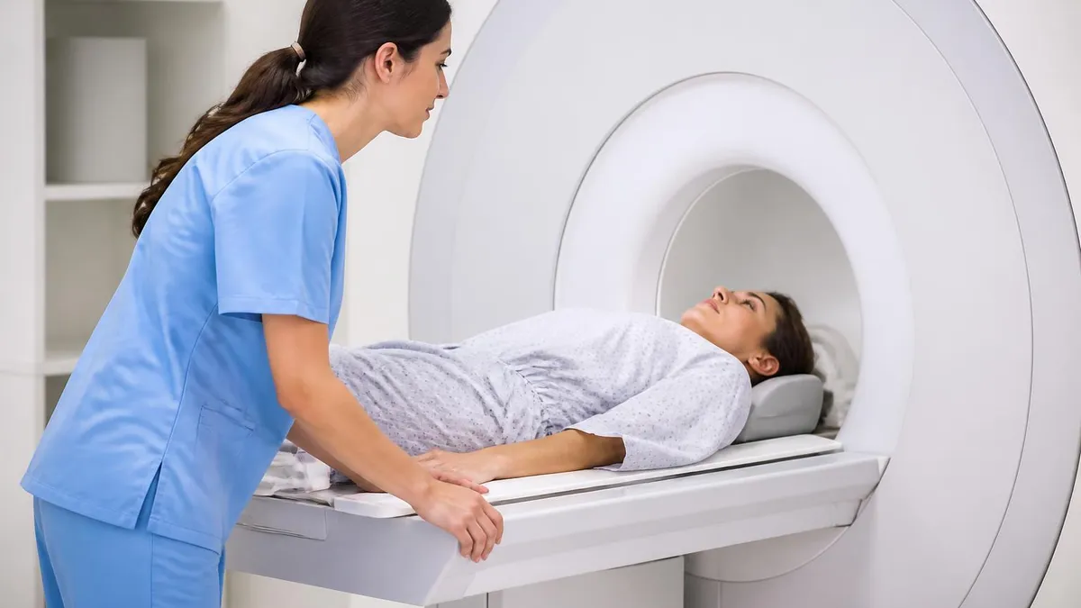
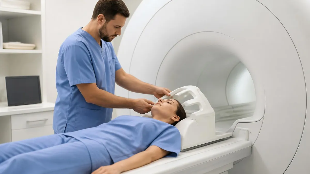
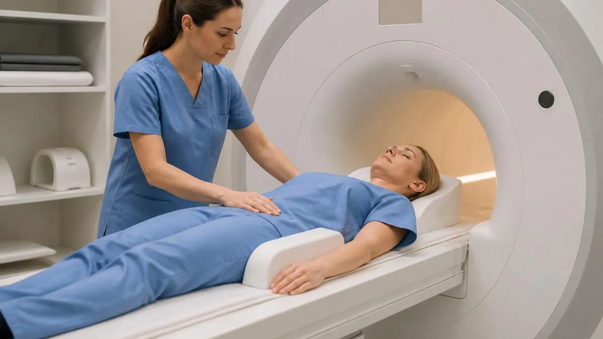
Patient Setup and Anesthesia
Probe Placement Under Real-Time MRI
Freeze-Thaw Cycle
Confirmation and Probe Removal
Recovery and Discharge
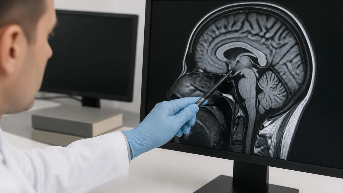
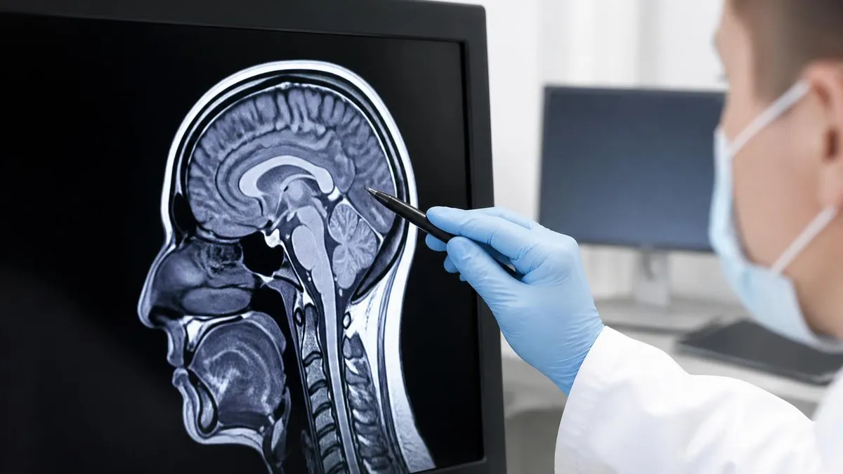
To understand how MRI guides cryoablation, you have to understand how MRI sees ice. Frozen tissue has essentially no mobile water protons, so it produces virtually no signal on any pulse sequence. The growing ice ball therefore appears as a sharply defined black void surrounded by normal-signal tissue, which is exactly what an interventionalist needs to see. The contrast is so striking that even a first-year radiology resident can identify the freeze margin within seconds of looking at the image.
The most commonly used sequence during active freezing is a fast spoiled gradient echo or balanced steady-state free precession acquisition, often with parallel imaging acceleration. These sequences provide images every two to three seconds with sufficient spatial resolution to track the ice ball as it expands by roughly two millimeters per minute. Many modern interventional suites also offer real-time thermometry using the proton resonance frequency shift method, which color-codes temperature directly onto the anatomic image. The full scope of MRI diagnostic capabilities extends well beyond static imaging into these interventional applications.
Cryoprobe selection is just as important as sequence selection. Standard stainless steel needles cause unacceptable susceptibility artifact at 1.5 Tesla or higher, blooming into halos several centimeters wide that obscure the very tissue you are trying to treat. Modern MRI-compatible probes use titanium-zirconium alloys with diameters between 1.5 and 2.4 millimeters. They produce a thin linear artifact that follows the probe shaft, leaving the surrounding tissue clearly visible. This engineering breakthrough is what made true real-time MRI guidance practical at clinical field strengths.
Coil selection also matters. A dedicated surface coil placed near the tumor improves signal-to-noise ratio dramatically compared to the integrated body coil, which is particularly important when imaging deep structures like the kidney or liver. Flexible loop coils with integrated cryoprobe pass-throughs are now commercially available, allowing the interventionalist to image and treat through the same anatomical window. Some institutions use a wide-bore 1.5T magnet specifically configured for interventional work, with a shortened bore length to improve patient access.
Two artifacts deserve special mention. First, the cryoprobe shaft can produce a small region of signal void beyond its tip on gradient echo sequences, which can be mistaken for early ice formation. Switching to a spin echo sequence eliminates this artifact and provides a true picture. Second, gas bubbles from rare equipment malfunctions can also appear as signal voids and may simulate ice ball extension; cross-checking with patient temperature monitoring and probe pressure readings prevents this misinterpretation.
For the technologist, the practical implications are clear. You must know which sequences your facility uses for planning, navigation, and confirmation. You must be able to position dedicated coils accurately and switch them out under sterile drapes if needed. You must recognize the difference between a properly forming ice ball and an artifact masquerading as one. And you must keep the table moving smoothly when the radiologist asks for the next slice — pauses during freezing can extend procedure time and compromise margin assessment.
The good news is that most of these skills transfer directly from diagnostic MRI experience. The unfamiliar element is the interventional workflow itself: working in a sterile field, communicating with anesthesia, and maintaining magnet safety with multiple metallic probes and gas lines in the room. With training, this becomes second nature, and many technologists report that interventional shifts are the most rewarding work in their careers.
MRI Practice Test Questions
Prepare for the MRI - Magnetic Resonance Imaging exam with our free practice test modules. Each quiz covers key topics to help you pass on your first try.
MRI Knowledge
MRI Exam Questions covering Knowledge. Master MRI Test concepts for certification prep.
MRI Physics
Free MRI Practice Test featuring Physics. Improve your MRI Exam score with mock test prep.
MRI Anatomy and Pathology
MRI Test Prep for MRI Anatomy and Pathology. Practice MRI Quiz questions and boost your score.
MRI Anatomy and Positioning
MRI Questions and Answers on MRI Anatomy and Positioning. Free MRI practice for exam readiness.
MRI Contrast Agents
Free MRI Quiz on MRI Contrast Agents. MRI Exam prep questions with detailed explanations.
MRI Patient Care and Positioning
MRI Practice Questions for MRI Patient Care and Positioning. Build confidence for your MRI certification exam.
Tumor Types Treated With MRI-Guided Cryoablation
Small renal masses under four centimeters, classified as T1a, are the most common indication. MRI-guided cryoablation achieves five-year local control rates of ninety-four percent in published series, comparable to partial nephrectomy without the surgical morbidity. The procedure preserves nephrons, which matters greatly for patients with solitary kidneys, chronic kidney disease, or bilateral tumors.
Tumor location relative to the collecting system, bowel, and renal vessels drives planning. Hydrodissection — instilling saline between the kidney and bowel — protects adjacent structures. Real-time MRI is particularly valuable here because the kidney moves with respiration and the safety margins are tight. Most patients are discharged within twenty-four hours with no change in baseline creatinine.
Is MRI-Guided Cryoablation Right for the Patient?
- +Outpatient procedure with same-day or next-day discharge
- +No surgical incision and minimal blood loss during treatment
- +Real-time visualization of the ice ball confirms complete tumor coverage
- +Repeatable if local recurrence occurs without scar tissue complications
- +Tissue preservation matters for kidneys, breasts, and weight-bearing bones
- +Excellent pain palliation for bone metastases within one week
- +Suitable for elderly or medically fragile patients who cannot tolerate surgery
- −Limited to tumors generally smaller than four centimeters in diameter
- −Requires specialized MRI-compatible probes and interventional suite
- −Higher upfront equipment cost than ultrasound or CT-guided alternatives
- −Longer procedure time than radiofrequency or microwave ablation
- −Not all insurers cover the procedure for every approved indication
- −Bleeding risk near vascular structures requires careful patient selection
- −Limited long-term data for some emerging indications like breast cancer
Technologist Preparation Checklist for MRI That Freezes Tumors
- ✓Verify patient screening form rules out implants incompatible with the interventional suite
- ✓Confirm MRI-compatible cryoprobes, hoses, and argon supply are in the room before patient arrival
- ✓Position dedicated surface coils on planned probe entry points and tape securely
- ✓Load planning images into the navigation workstation and confirm DICOM linkage
- ✓Set up real-time fast gradient echo sequence with parallel imaging acceleration
- ✓Establish sterile field with MRI-safe drapes covering the bore opening
- ✓Place MRI-conditional pulse oximeter, ECG, and blood pressure cuff on patient
- ✓Confirm anesthesia gas lines and monitors are routed safely outside the five-gauss line
- ✓Brief the team on emergency quench procedures and code response in the magnet room
- ✓Time freeze and thaw cycles precisely and document on the procedure record
The five-millimeter margin rule is non-negotiable
Studies show that local recurrence rates rise sharply when the ice ball extends less than five millimeters beyond the tumor edge in any plane. Always confirm the margin in three orthogonal views before terminating the second freeze cycle. The ice ball periphery is at zero degrees Celsius, but cytotoxic temperatures only reach roughly five millimeters inside that visible edge.
The evidence base for MRI-guided cryoablation has matured substantially over the past fifteen years, with multi-center registries, single-arm prospective trials, and a growing number of randomized comparisons against surgery and other ablative modalities. The strongest data come from renal cell carcinoma, where five-year local recurrence-free survival rates of ninety-four percent and cancer-specific survival rates above ninety-seven percent for T1a tumors are now well established in the peer-reviewed literature.
Complication rates are notably low. Major complications occur in two to four percent of cases, with bleeding requiring transfusion being the most common and typically managed with embolization rather than open surgery. Pneumothorax can occur during transthoracic approaches and is treated with a small-bore chest tube. Urinary fistula, bowel injury, and nerve damage are rare but documented, almost always associated with tumors in challenging locations that pushed the limits of safe margins.
Pain outcomes deserve special attention. For bone metastases, pain scores measured on validated instruments like the Brief Pain Inventory drop by fifty to seventy percent within one week and remain durable through six months in most patients. This effect is dramatic and reproducible, which is why national guidelines now list cryoablation as a first-line option for refractory cancer pain in selected sites, ahead of repeat radiation in many cases.
The patient experience is generally favorable. Quality-of-life questionnaires administered at one month show return-to-baseline scores for the majority of patients, compared to several months for surgical patients. Cosmetic outcomes are excellent because the only skin marks are the small probe entry points, which heal as nearly invisible scars. Body image and emotional recovery, particularly for breast and prostate cases, are meaningfully better than after major surgery.
Cost-effectiveness analyses generally favor cryoablation over surgery for appropriate candidates. Although upfront procedural costs can be comparable, shorter hospital stays, faster return to work, and lower complication rates produce net savings for the healthcare system. Some payers still classify the procedure as experimental for certain indications, so verifying coverage during pre-authorization is an important workflow step that should not be left to the last minute.
For technologists, knowing this outcome data has two practical benefits. First, you can answer patient questions accurately during the screening interview, which builds trust and helps patients feel informed. Second, when a procedure runs long or encounters difficulty, you understand what is at stake and why the radiologist insists on getting that margin right. Procedures done well produce decades of disease control; procedures rushed or compromised produce recurrence and reoperation.
One area to watch closely is the expanding role of cryoablation in breast cancer. The ICE3 trial reported encouraging interim data for elderly women with small invasive cancers, and randomized trials against lumpectomy are ongoing. If these trials show non-inferiority, MRI-guided cryoablation could substantially change the landscape of early breast cancer treatment over the next decade.
Argon and helium gas lines entering the magnet room must be secured against projectile risk and quench-related pressure changes. Never leave gas bottles unsecured near the bore. Verify that all cryoprobes, monitoring leads, and warming catheters are explicitly labeled MRI-conditional for your specific field strength. A single ferromagnetic instrument can cause catastrophic injury at 1.5 or 3.0 Tesla.
When comparing MRI-guided cryoablation to alternatives, the most useful framework is to match the modality to the clinical question. For a small renal mass in a patient with one kidney and chronic kidney disease, cryoablation under MRI guidance is often superior to partial nephrectomy because it preserves more nephrons and avoids ischemia time. For a healthy patient with the same tumor but two kidneys, partial nephrectomy may offer marginally better long-term outcomes that justify the bigger surgery.
Radiofrequency ablation, which uses heat instead of cold, has a longer track record and lower equipment cost but is harder to monitor in real time. The heated tissue does not produce the same crisp imaging signature as ice. Microwave ablation creates larger zones faster but with less predictable shapes. Cryoablation wins on visualization and shape control; the heat-based modalities win on speed and ablation volume for larger tumors. Many centers use both, choosing the tool that fits the case.
CT-guided cryoablation remains the most common platform in the United States simply because CT suites are more available and cheaper to operate. The trade-off is reduced soft tissue contrast and difficulty seeing the ice ball margin precisely. For tumors abutting critical structures like nerves, bowel, or the spinal canal, MRI guidance is the better choice and is often the only choice that meets standard of care for safety.
A growing number of interventional radiologists are pursuing fellowship training that includes both platforms. For those considering this career path, our guide on how to become an MRI technician describes the foundational training that opens doors to interventional roles.
Stereotactic body radiation therapy is another competitor, particularly for inoperable lung and liver tumors. It is non-invasive but requires multiple treatment sessions and carries radiation-related risks. Cryoablation completes treatment in one session and produces no ionizing radiation exposure, which matters for younger patients and those with prior radiotherapy who cannot receive additional dose to the same area.
For benign conditions, the calculus is different. A benign fibroadenoma does not need to be removed at all in most cases, so the comparison is between surveillance and a single brief intervention. Cryoablation offers a definitive treatment that produces no scar and gradually eliminates the palpable mass, which many women prefer over years of repeat imaging surveillance. Patient preference, more than survival data, drives this decision.
Cost varies widely by region, payer, and indication. Self-pay costs for MRI-guided renal cryoablation in the United States typically range from twenty to thirty-five thousand dollars all-in, including pre-procedure imaging, anesthesia, the procedure itself, and post-procedure surveillance. Medicare and most commercial insurers cover the procedure for established indications when documentation supports medical necessity. Pre-authorization is essential, and denials are common for newer indications that lack the depth of evidence required by some plans.
The bottom line for patients and referring physicians is that MRI-guided cryoablation is not the right answer for every tumor, but it is increasingly the right answer for a meaningful subset where tissue preservation, imaging precision, and rapid recovery all matter. Knowing when to recommend it — and when to recommend something else — is the mark of mature clinical judgment.
Practical tips for technologists and trainees preparing to work in an MRI-guided cryoablation program begin with anatomy. You will not be effective in this role if you cannot quickly identify the renal hilum, the celiac trunk, the brachial plexus, or the conus medullaris on axial, coronal, and sagittal sections. Spend time before your first interventional shift reviewing cross-sectional anatomy with an emphasis on the regions your service treats most often. The radiologist will move quickly, and you need to keep pace.
Second, learn your scanner's interactive imaging mode inside and out. Most major vendors offer a real-time mode that allows the operator to update slice position with a trackball or controller without re-prescribing the full sequence. Practice during downtime so that during a live procedure you can adjust angulation, change contrast weighting, and zoom in on the ice ball margin without fumbling. Smooth scanner control is the difference between a confident team and a chaotic one.
Third, develop a mental checklist for the moment just before freezing begins. Confirm probe positions on three planes. Verify that the dedicated surface coil is taped down and will not shift. Note the time. Set up the next sequence in the queue so you can switch instantly if the radiologist requests a different contrast. Check that the argon supply pressure is in range and that the room temperature monitor is on screen. These thirty seconds of preparation prevent ninety percent of intra-procedural disruptions.
Fourth, communicate clearly and continuously. Interventional MRI is a team sport involving the radiologist, anesthesia, nursing, and you. Call out time elapsed in the freeze cycle every two minutes. Announce when you are starting a new sequence so the radiologist does not look up at a blank screen. If you see an artifact or a change you do not understand, say so immediately — silence does not equal competence in this setting.
Fifth, document meticulously. Procedure times, probe types and serial numbers, sequence parameters, contrast agent doses, and any complications must all be recorded contemporaneously. This documentation matters for billing, quality improvement, malpractice protection, and patient safety. Many programs use a structured intra-procedural template; if yours does not, advocate for one and offer to help build it.
Sixth, debrief after every case. The team should spend three to five minutes after each procedure reviewing what went well and what could improve. This discipline accelerates learning faster than any formal course and builds the team trust that complex procedures demand. Junior technologists who participate actively in debriefs progress to lead-tech status faster than those who simply complete their tasks and leave.
Finally, keep learning. Subscribe to journals like the Journal of Vascular and Interventional Radiology and Radiology, attend at least one national meeting per year if possible, and seek out hands-on workshops. The field is evolving rapidly, and the technologists who thrive are those who stay current. Within five years, expect to see new probe designs, AI-assisted planning tools, and expanded indications that will reshape how you do this work day to day.
MRI Questions and Answers
About the Author
Medical Laboratory Scientist & Clinical Certification Expert
Johns Hopkins UniversityDr. Sandra Kim holds a PhD in Clinical Laboratory Science from Johns Hopkins University and is certified as a Medical Technologist (MT) and Medical Laboratory Scientist (MLS) through ASCP. With 16 years of clinical laboratory experience spanning hematology, microbiology, and molecular diagnostics, she prepares candidates for ASCP board exams, MLT, MLS, and specialist certification tests.
Join the Discussion
Connect with other students preparing for this exam. Share tips, ask questions, and get advice from people who have been there.
View discussion (6 replies)