Xience Stent and MRI Safety: A Complete Patient and Technologist Guide 2026 July
🎓 Xience stent and MRI safety explained: scan timing, field strength limits, screening rules, and what every patient and technologist must verify.
The topic of xience stent and mri safety comes up almost every day in modern imaging departments, and for good reason. Coronary stents are among the most common cardiovascular implants in the United States, with hundreds of thousands placed each year, and many of those patients eventually need magnetic resonance imaging for unrelated reasons. Understanding when an MRI can be performed safely, what field strengths are acceptable, and what documentation must be reviewed first protects patients from real harm while preventing unnecessary scan cancellations.
Xience drug-eluting stents are manufactured by Abbott Vascular and are built on a cobalt-chromium platform that is officially labeled MR Conditional. That designation does not mean unsafe. It means safe under specific, documented conditions. The exact conditions matter, and they differ slightly depending on the Xience generation, the implant date, and the magnet strength of the MRI scanner. Technologists who memorize one rule and apply it to every cardiac patient will eventually meet a case where that rule is wrong.
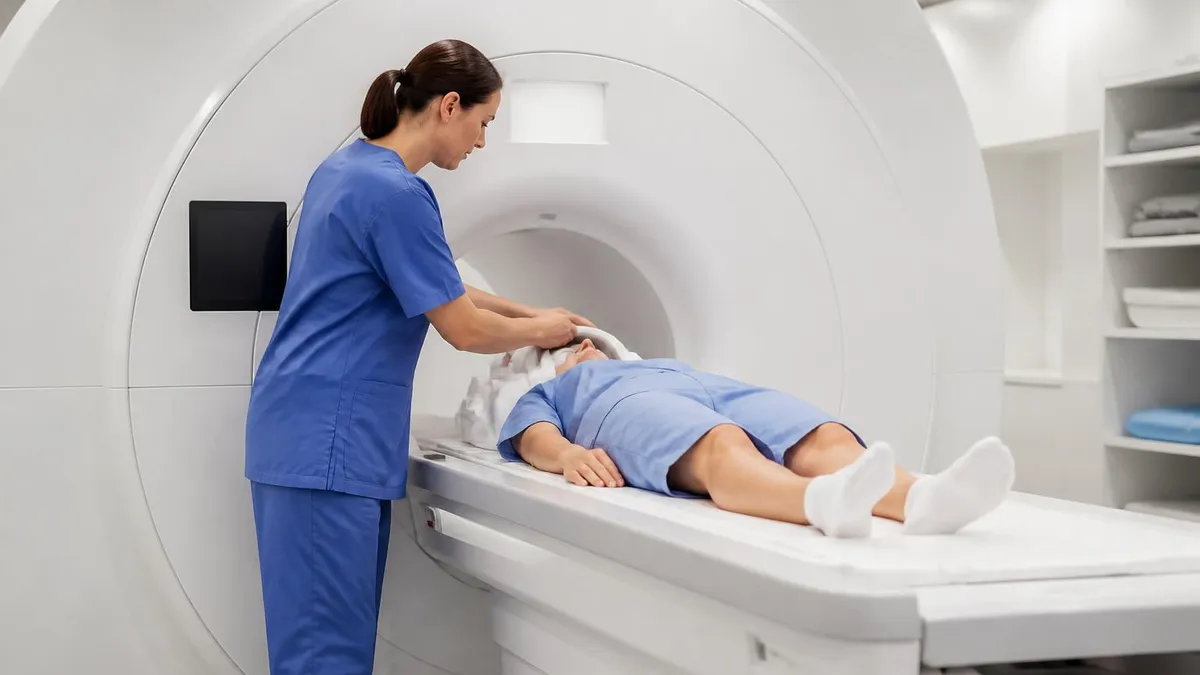
This guide walks through everything a patient, referring clinician, or MRI technologist needs to confirm before sending a Xience stent recipient into the bore. We cover the waiting period myth, the truth about static field limits, the role of specific absorption rate, what to do when documentation is missing, and how to handle patients who have stents stacked alongside pacemakers or aneurysm clips. The goal is to leave you confident in your screening workflow, not anxious about every cardiac history line.
We also clarify common patient questions that come up at the front desk, like whether the stent can be pulled out of place by the magnet, whether contrast is dangerous with a metal implant, and whether a smartwatch or fitness tracker interferes with anything. Spoiler: the engineering behind cobalt-chromium alloys is more reassuring than most patients expect, but the screening workflow still demands precision.
Finally, this article addresses the registry and board exam angle. Technologists preparing for the ARRT MRI registry or the ARMRIT exam will see stent safety questions, and they often hinge on small distinctions like 1.5T versus 3T conditional labeling, the meaning of MR Conditional symbols, and the difference between ferromagnetic and weakly paramagnetic materials. Memorizing the right rule once saves time on exam day and shift after shift in clinical practice.
By the end of this read, you should be able to look at a patient history mentioning a Xience V, Xience Xpedition, Xience Sierra, or Xience Skypoint stent and know exactly what your next screening step is. You should also understand why the old six-week waiting period no longer applies to most modern stents, and why scanning too soon was never actually the catastrophic risk many earlier safety posters suggested.
Xience Stents and MRI Safety by the Numbers
Xience Stent Generations and MR Status
The original everolimus-eluting stent on a cobalt-chromium L605 platform. MR Conditional at 1.5T and 3.0T immediately after implantation per Abbott's instructions for use.
Second-generation refinements with improved deliverability and the same cobalt-chromium alloy. Identical MR Conditional labeling, with no required waiting period before clinically indicated MRI.
Released with an enhanced low-profile delivery system. Maintains the same MR Conditional safety profile at 1.5T and 3.0T under standard SAR limits for normal operating mode.
Latest generations with expanded size matrices for complex lesions. Both are MR Conditional immediately post-implantation and have been independently tested at clinical field strengths.
All Xience stents ship with an implant card that lists the model, length, diameter, and MR conditional language. Patients should keep this card and present it at every imaging visit.
Understanding why a Xience stent is safe in the MRI environment requires a quick tour of the physics involved. The static magnetic field of an MRI scanner is enormously strong, with 1.5T magnets producing about 30,000 times the Earth's field and 3T magnets doubling that. Yet field strength alone is not what determines whether an implant is safe. The deciding factors are the magnetic susceptibility of the implant material, its geometry, how firmly it is anchored in tissue, and how the scanner couples energy into it.
Cobalt-chromium L605, the alloy used in every modern Xience stent, is classified as weakly paramagnetic. That means it experiences a tiny attraction to the magnet but nothing remotely close to the violent pull that ferromagnetic materials like older surgical steel can exhibit. In bench testing, the magnetically induced displacement force on a Xience stent is a small fraction of the gravitational force on the device itself, so the bore's pull cannot move it. The torque applied by the gradient fields is similarly small.
The more nuanced concern is radiofrequency heating. When the MRI's RF coil transmits, electrically conductive implants can absorb energy and warm slightly. Bench and modeling studies on Xience stents at both 1.5T and 3T have shown peak temperature increases well under the threshold considered clinically significant, typically a fraction of a degree under standard operating conditions. The endothelium that grows over the stent struts during the first month also acts as a thermal buffer, but even bare stents have not produced harmful heating in validated tests.
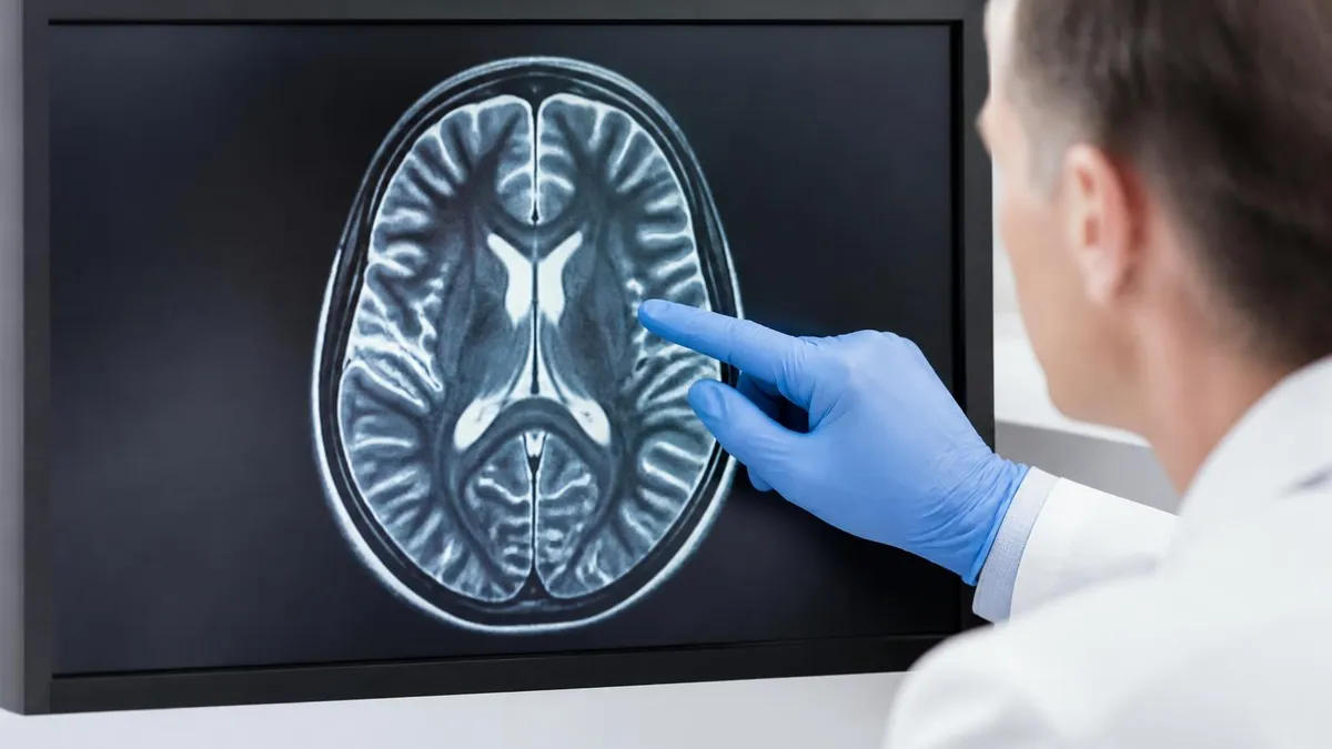
The third interaction is image artifact. Coronary stents create local signal voids and geometric distortion roughly proportional to the strut thickness and stent diameter. For a typical Xience strut at 81 micrometers, the artifact is small compared with older bare-metal designs and is rarely a barrier to reading non-cardiac MRI. For coronary MR angiography directly through the stent lumen, the artifact still limits diagnostic yield, which is why most cardiologists prefer CT angiography for in-stent restenosis assessment.
It is worth contrasting Xience with implants that genuinely require caution. A retained shrapnel fragment, an older cerebral aneurysm clip made from martensitic stainless steel, or certain cochlear implants behave very differently in the bore. Stent technology evolved precisely to avoid these problems, with materials chosen as much for MRI compatibility as for biological performance.
Patients often ask whether subsequent imaging beyond MRI is affected. The answer is no. CT scans, ultrasound, fluoroscopy, and nuclear medicine studies have no meaningful interaction with the stent. Even MRI of regions far from the chest, such as a knee or brain, is unaffected by the stent's presence in any practical way because the local artifact stays localized.
The takeaway from the physics is straightforward. The Xience platform was engineered with magnetic compatibility in mind, the safety data is mature, and the screening process is now about confirming the right model and following standard SAR rules rather than worrying about whether the scan can be done at all.
MRI Practice Test Questions
Prepare for the MRI - Magnetic Resonance Imaging exam with our free practice test modules. Each quiz covers key topics to help you pass on your first try.
MRI Knowledge
MRI Exam Questions covering Knowledge. Master MRI Test concepts for certification prep.
MRI Physics
Free MRI Practice Test featuring Physics. Improve your MRI Exam score with mock test prep.
MRI Anatomy and Pathology
MRI Test Prep for MRI Anatomy and Pathology. Practice MRI Quiz questions and boost your score.
MRI Anatomy and Positioning
MRI Questions and Answers on MRI Anatomy and Positioning. Free MRI practice for exam readiness.
MRI Contrast Agents
Free MRI Quiz on MRI Contrast Agents. MRI Exam prep questions with detailed explanations.
MRI Patient Care and Positioning
MRI Practice Questions for MRI Patient Care and Positioning. Build confidence for your MRI certification exam.
Field Strength, Timing, and SAR Rules for Xience Stent and MRI Safety
Every modern Xience stent is labeled MR Conditional at both 1.5 Tesla and 3.0 Tesla. That covers the vast majority of clinical MRI scanners installed in the United States. Higher field research scanners at 7T are not covered by Abbott's labeling and are typically restricted to specific approved research protocols. If your facility uses a 1.5T or 3T magnet, the field strength itself is not a barrier to scanning a Xience recipient.
What technologists must verify is that the labeling applies to the specific implant. The patient's implant card, the operative report, or the cath lab note will confirm the model. If the card is missing and the patient cannot recall details, a quick call to the implanting facility or the patient's cardiologist usually resolves the question within minutes.
Scanning a Xience Stent Patient: Practical Pros and Cons
- +No waiting period required after stent placement for clinically indicated MRI
- +MR Conditional at both 1.5T and 3.0T, covering nearly all US clinical scanners
- +Cobalt-chromium alloy is weakly paramagnetic with negligible displacement force
- +RF heating remains well below clinically significant thresholds under normal SAR
- +Local artifact stays confined and rarely interferes with non-cardiac imaging
- +Implant cards make documentation and verification straightforward
- +Standard screening workflow applies, no special coils or protocols required
- −Coronary MR angiography through the stent lumen has limited diagnostic value
- −Missing implant cards force time-consuming verification calls before scanning
- −First-level SAR modes and research protocols may exceed conditional limits
- −Patients sometimes confuse Xience with older bare-metal stents and panic
- −Older Xience V cards from 2008 era may lack updated MR language
- −Pacemaker or aneurysm clip coexisting with stent multiplies screening complexity
Pre-Scan Checklist for Xience Stent and MRI Safety
- ✓Confirm the implant manufacturer and model on the implant card or operative note
- ✓Verify the stent is a Xience generation, not a different drug-eluting or bare-metal device
- ✓Note the implantation date and any other cardiac devices placed at the same procedure
- ✓Check that scanner field strength is 1.5T or 3.0T as labeled
- ✓Set scanner to normal operating mode with whole-body SAR at or below 2.0 W/kg
- ✓Screen for additional implants such as pacemakers, ICDs, or aneurysm clips
- ✓Review renal function if gadolinium contrast is planned for cardiac or vascular MRI
- ✓Document patient symptoms before the scan as a baseline reference
- ✓Confirm patient understanding and obtain informed consent per facility policy
- ✓Brief the radiologist about the stent location so artifact is anticipated on review
No waiting period is required for modern Xience stents
Abbott's instructions for use explicitly allow MRI immediately after Xience stent implantation under standard 1.5T or 3.0T conditional parameters. The old six-week rule belongs to first-generation bare-metal stents and should not delay urgent imaging. When in doubt, refer to the implant card or contact the implanting cardiologist for fast confirmation.
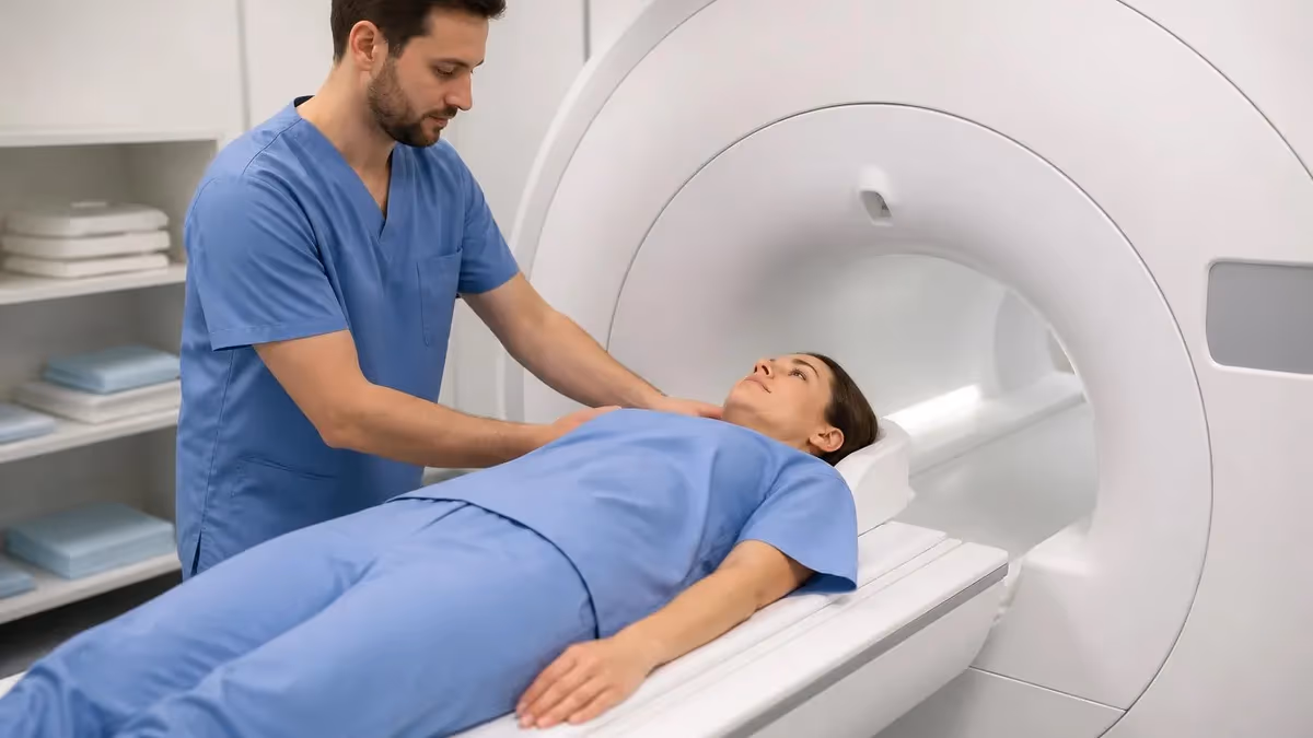
Communicating with a patient who has a Xience stent before an MRI is half the safety equation. Many people who received a stent during an acute coronary event remember very little about the device itself. They may not know the brand, the model, or even whether more than one stent was placed. A calm, structured conversation that explains why you are asking these questions is far more effective than a checklist read in a hurry.
Start by acknowledging that having a cardiac stent does not disqualify them from MRI. That single sentence relieves a lot of anxiety. Then ask whether they have an implant card, which is the small wallet-sized document that lists the device specifications. If they do not, ask whether the stent was placed within the last twenty years and at which hospital. Most modern stents placed in the US since the mid-2000s are MR Conditional, but verification is still required.
Patients often ask whether the magnet can move the stent or pull it out of the artery. Reassure them that the cobalt-chromium alloy used in Xience devices is not magnetic in the everyday sense. The stent is also fully endothelialized within a few weeks, meaning a layer of vascular tissue has grown over and through the struts, locking it permanently in place. Even in the immediate post-implant window before endothelialization, the displacement force from a clinical magnet is far too small to dislodge it.
Heating is the next common worry. Explain that bench testing and clinical experience demonstrate any temperature rise during a normal MRI is a fraction of a degree, well below the threshold for any tissue injury. Patients may also ask about the contrast agent. Gadolinium-based contrast does not interact with the stent. The only contrast consideration is the patient's kidney function, which the radiologist already screens for before any gadolinium administration.
If the patient has more than one implant, the conversation expands. Coexisting pacemakers, defibrillators, deep brain stimulators, or older aneurysm clips each have their own screening rules, and those rules take priority over the stent. The stent itself rarely changes the decision, but the other devices may. In that situation, an MRI safety officer or radiologist should review the full implant inventory before the scan proceeds.
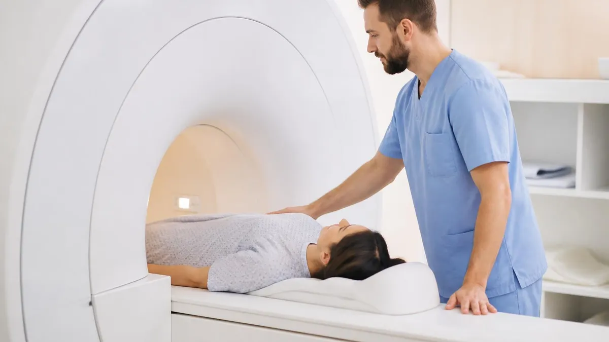
Finally, walk the patient through what they will experience during the scan. Loud knocking sounds, table movement, the requirement to stay still, and the breath-hold instructions are unchanged because of the stent. They can wear the call button, communicate with the technologist, and stop the scan at any time if they feel unwell. Many patients are surprised that having a stent does not make the procedure feel any different from a routine MRI.
Documentation closes the loop. Record the model, the implantation date, the field strength used, the SAR mode, the duration of the scan, and any patient-reported sensations during and after the procedure. That record helps if the patient returns for another MRI later, and it builds an institutional baseline that future technologists can rely on.

Never rely on a patient's verbal recollection alone when a stent was placed more than ten years ago. Older bare-metal stents and certain non-Xience devices have different conditional parameters or, in rare cases, are not MR Conditional. Confirm the model in writing through the implant card, the operative report, or a call to the implanting cardiologist before the patient enters the bore.
For technologists preparing for the ARRT MRI registry, the ARMRIT exam, or in-house competency testing, stent questions are a frequent topic. The exam writers know that real-world screening errors often stem from a misunderstanding of MR Conditional labeling, so they test that distinction directly. Knowing that Xience is MR Conditional rather than MR Safe is the first nuance to internalize. MR Safe means the implant poses no known hazard in any MRI environment, while MR Conditional means it is safe only under documented conditions.
A second exam favorite is the difference between field strength labeling and SAR labeling. A stent can be approved at 1.5T and 3.0T but still require normal operating mode for SAR. Confusing the two leads to wrong answers. The labeling reads as a stack of conditions that all must be satisfied, not a single threshold. Practice reading vendor labels carefully and parsing them into separate parameters.
Material classification is the third recurring theme. Cobalt-chromium L605 is weakly paramagnetic. Nitinol, used in some peripheral and neurovascular devices, is also non-ferromagnetic but has its own thermal characteristics. Older stainless steel stents, particularly 316L, are also classified as MR Conditional under most modern labeling. Knowing that ferromagnetic materials in older implants are the actual safety concern, not modern cardiovascular stents, anchors the broader topic in your memory.
Exam questions also test the timing myth. If a question asks how long after Xience placement an MRI can be performed, the correct answer per manufacturer labeling is immediately when clinically indicated. Older study materials still list six weeks. Trust the current manufacturer documentation, not outdated lecture notes. When real exam answer choices include both, the manufacturer-aligned answer is the correct one.
Scenario-based questions tie everything together. A typical stem might describe a patient three weeks post Xience implantation needing an MRI for suspected stroke. The correct workflow is to proceed under standard MR Conditional parameters at 1.5T or 3.0T in normal operating mode after verifying the implant. Distractors will suggest waiting, lowering the field strength, or canceling the scan, all of which are wrong.
Finally, watch for combination implants on the exam. A patient with a Xience stent and a pacemaker will be evaluated based on the more restrictive device. The stent itself remains scannable, but the pacemaker drives the protocol. Know how to identify which device is the limiting factor, and remember that you should escalate to the radiologist or MR safety officer when implants overlap in unusual ways.
Build your study around real labels, real screening forms, and real scenarios. Memorizing isolated facts about stents rarely helps in clinical practice or on exam day. Understanding why each rule exists transforms a list of trivia into a reliable framework you can apply to any new implant you encounter, including ones that did not exist when your textbook was printed.
Practical preparation for stent safety screening is built on small habits that prevent the rare but serious errors that make headlines. Keep a printed quick-reference card at the screening station listing the major US stent manufacturers, their MR Conditional parameters, and the phone numbers for their safety information lines. Abbott, Boston Scientific, Medtronic, and Cordis all publish technical service numbers that respond within minutes to verification questions, and saving those contacts saves hours of delay.
Maintain a department logbook of unusual cases. When a patient arrives with an unfamiliar device or an out-of-country stent that does not appear in your usual reference, document how the case was resolved and what source confirmed the safety. The log becomes an institutional memory that helps the next technologist who encounters the same device on a busy weekend shift with no radiologist on site.
Run drills on missing-documentation scenarios. Practice the workflow of contacting the implanting facility, pulling old operative reports through your health information exchange, and escalating to the on-call radiologist when the device cannot be confirmed. The first time you have to do this should not be when a patient is already on the table. Build the muscle memory in calm conditions.
Refresh your knowledge of the MR Conditional symbol itself. The official IEC-defined symbol shows the letters MR inside a triangle with the word Conditional, sometimes accompanied by specific parameters. Recognizing the symbol on packaging and implant cards speeds up the verification process. Train all front-desk staff to recognize it too so they can route patients to the MRI safety officer immediately when an implant card is presented.
Coordinate with the cardiology and electrophysiology services. The patients with the most complex implant histories tend to receive their devices in those departments, and an early conversation about screening expectations prevents misunderstandings. Some institutions go further and tag patients in the electronic health record with their stent details so the information populates automatically on every imaging order.
Educate patients to be their own advocates. Tell them to keep their implant card in their wallet, photograph it as a backup on their phone, and present it at every imaging visit. A patient who arrives with documentation in hand cuts your screening time dramatically and reduces the risk of error. Many hospital systems now also provide a digital implant passport that lives in the patient portal for easy retrieval.
Finally, stay current. MRI safety guidance evolves as new implants come to market and as research clarifies older devices. The ACR Manual on MR Safety updates regularly, the manufacturer instructions for use are revised when new testing results in updated labeling, and professional societies publish position papers that change everyday practice. A standing quarterly review of these sources keeps your department's protocols aligned with the best available evidence.
MRI Questions and Answers


About the Author
Medical Laboratory Scientist & Clinical Certification Expert
Johns Hopkins UniversityDr. Sandra Kim holds a PhD in Clinical Laboratory Science from Johns Hopkins University and is certified as a Medical Technologist (MT) and Medical Laboratory Scientist (MLS) through ASCP. With 16 years of clinical laboratory experience spanning hematology, microbiology, and molecular diagnostics, she prepares candidates for ASCP board exams, MLT, MLS, and specialist certification tests.
Join the Discussion
Connect with other students preparing for this exam. Share tips, ask questions, and get advice from people who have been there.
View discussion (6 replies)