MRI Scan of Brain: How It Works, What It Shows, and What to Expect 2026 July
An MRI scan of brain explained: ✏️ how the machine works, what it detects, prep tips, sequences, safety, and how to prepare for MRI exams and registry tests.
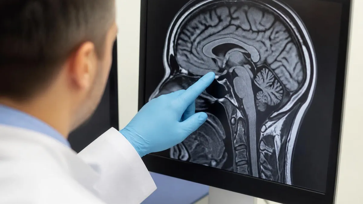
An MRI scan of brain is one of the most powerful diagnostic tools in modern medicine, producing detailed cross-sectional images of soft tissue without using ionizing radiation. Unlike a CT scan, which relies on X-rays, magnetic resonance imaging uses a strong magnetic field and radiofrequency pulses to map the hydrogen atoms inside your body. Because the brain is rich in water and fat, both packed with hydrogen, it shows up with remarkable clarity, letting radiologists see structures as small as a few millimeters across.
When a physician orders an MRI scan of brain, they are usually looking for answers about headaches, seizures, vision changes, memory loss, or suspected stroke. The scan can reveal tumors, multiple sclerosis plaques, aneurysms, bleeding, infection, and developmental abnormalities long before they would appear on other imaging. For students preparing for MRI registry exams, understanding why the brain images so well is the foundation for nearly every clinical and physics question you will encounter on test day.
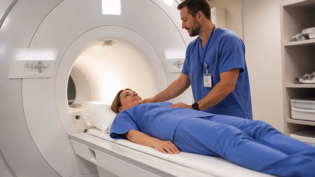
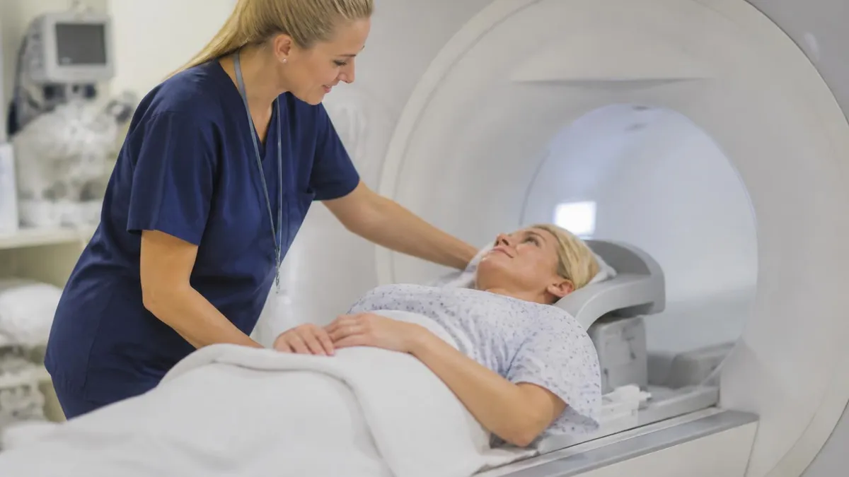
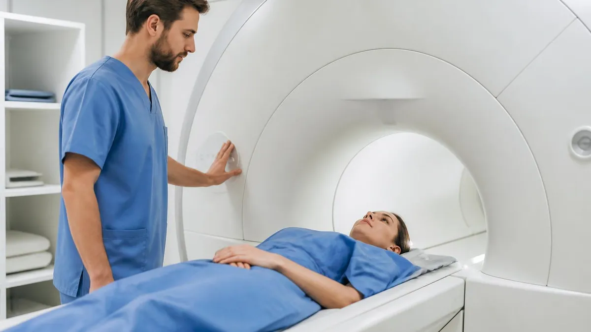
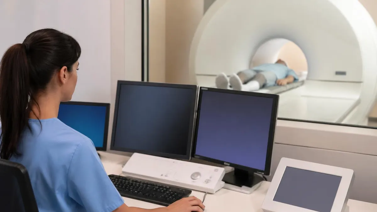
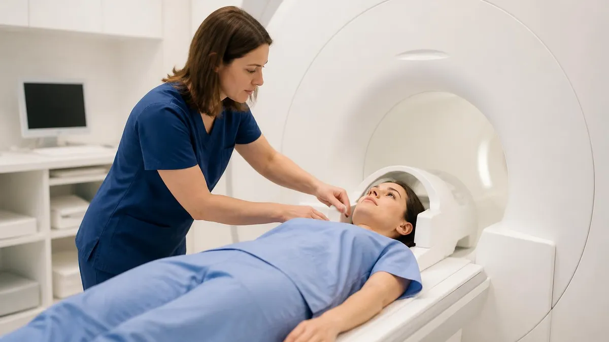
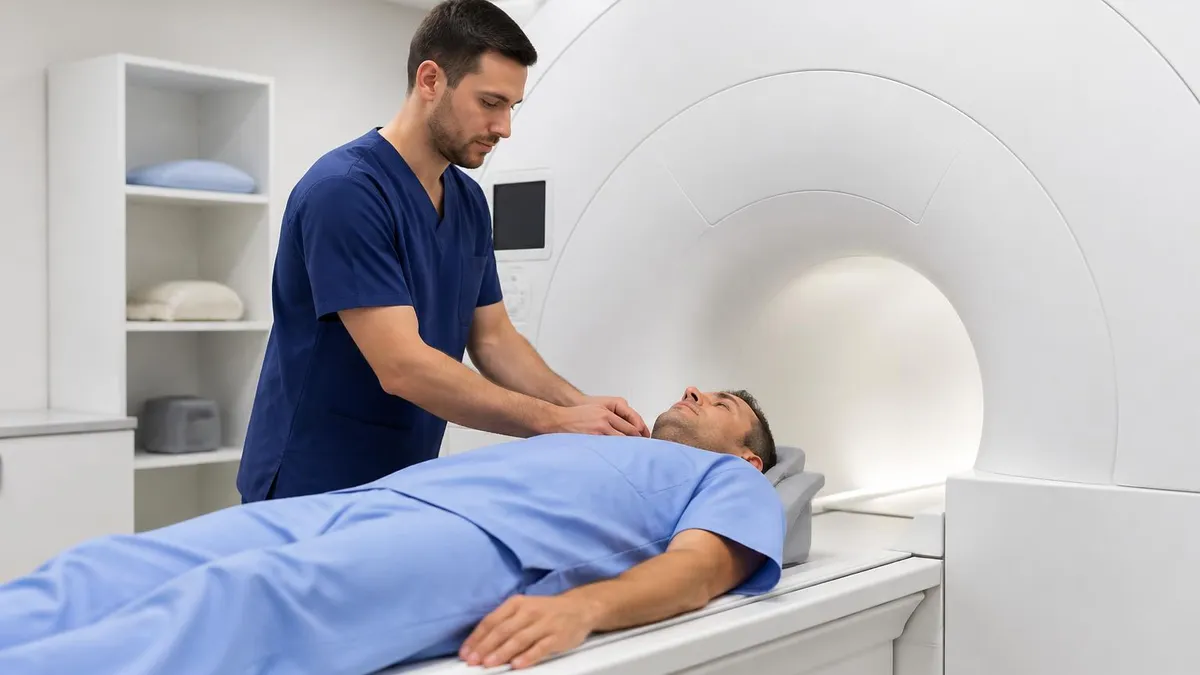
The procedure itself is painless and typically lasts between 30 and 60 minutes. You lie on a motorized table that slides into a cylindrical magnet, and a coil is placed around your head to receive the radio signals. The machine produces loud knocking and buzzing sounds as gradient coils switch on and off, which is why patients are given earplugs or headphones. Staying still is critical, because even slight movement blurs the final images and can force a repeat sequence.
Radiologists rarely rely on a single image. A brain study includes multiple sequences, each weighted to highlight different tissue properties. T1-weighted images show anatomy beautifully, T2-weighted images make fluid and edema bright, and FLAIR sequences suppress cerebrospinal fluid so that subtle lesions stand out. Diffusion-weighted imaging is the workhorse for detecting acute stroke within minutes of symptom onset, making it indispensable in emergency neurology and a frequent topic on board questions.
If you want to see what a textbook-perfect result looks like before studying disease states, comparing your knowledge against a mri scan of brain that is entirely normal builds the visual baseline every technologist needs. Recognizing the gray-white matter junction, the ventricular system, and symmetric basal ganglia trains your eye to spot when something is wrong, which is precisely the skill examiners test.
This guide walks through how the scan works, what each sequence reveals, how to prepare patients safely, and how to study the material for certification. Whether you are a patient facing your first appointment, a student cramming for the ARRT MRI registry, or a technologist refreshing protocols, the sections below break the topic into clear, practical pieces. By the end you will understand not just the what, but the why behind every brain MRI you encounter.
Brain MRI by the Numbers
How a Brain MRI Actually Works
Inside the bore, the strong static magnetic field aligns the hydrogen protons in your brain tissue along its axis. The vast majority cancel out, but a tiny net surplus creates the magnetization that imaging exploits.
A radiofrequency pulse tuned to the Larmor frequency tips the aligned protons out of equilibrium. This is the moment energy is absorbed, knocking the magnetization into the transverse plane where it can be measured by the coil.
When the pulse stops, protons release energy and realign. The rates of this recovery, called T1 and T2 relaxation, differ by tissue type and produce the contrast that distinguishes gray matter, white matter, and fluid.
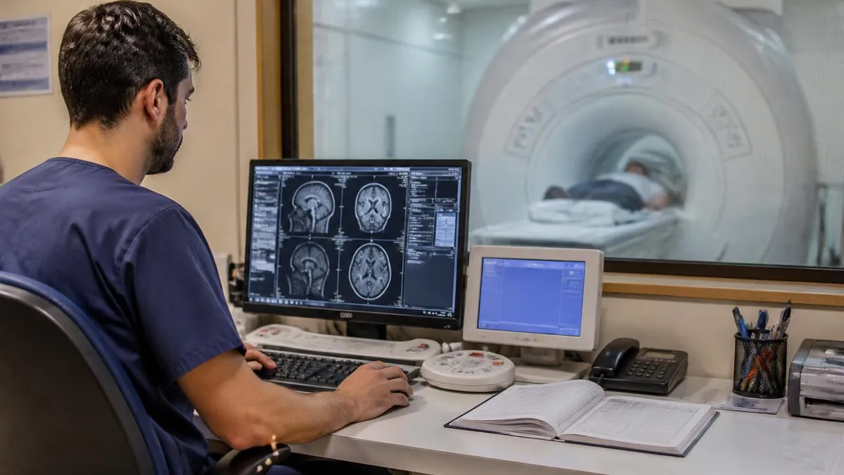
Gradient coils create controlled variations in the field so each location resonates at a slightly different frequency. The scanner uses this spatial encoding and a Fourier transform to reconstruct a precise image slice.
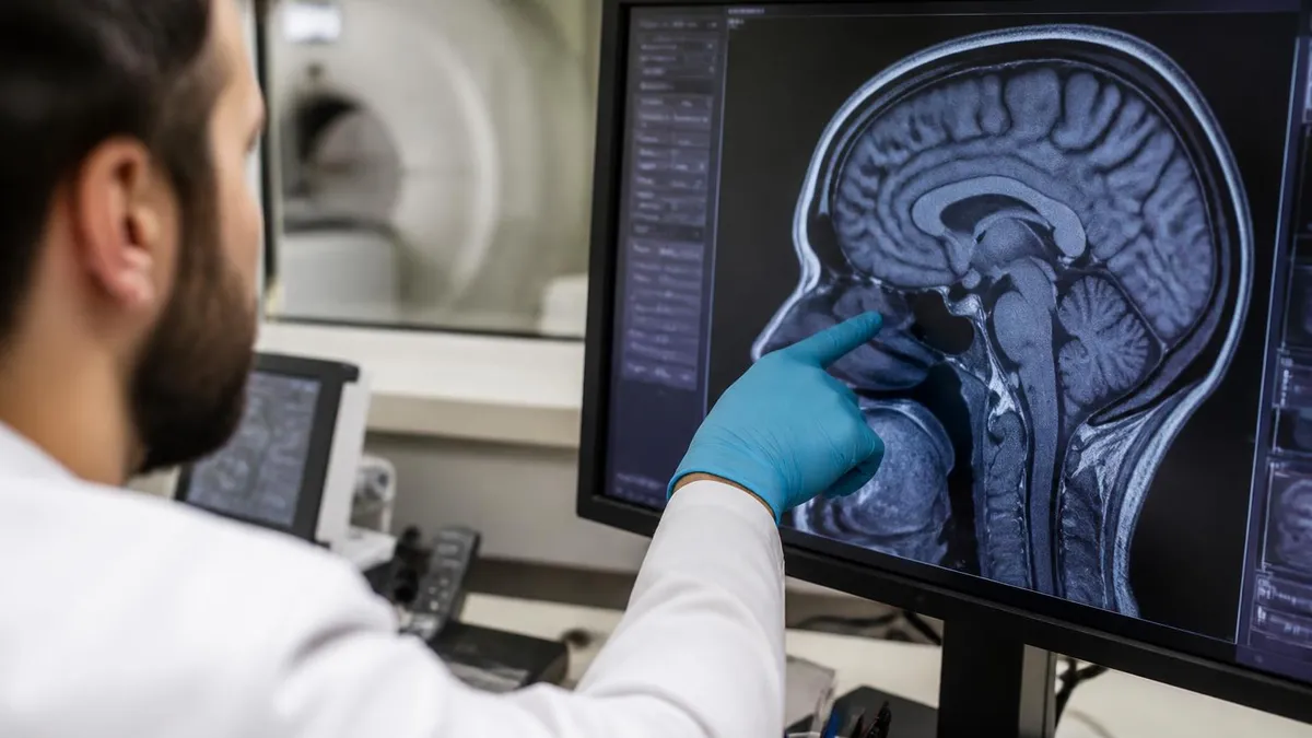
What makes an MRI scan of brain so clinically valuable is its unmatched soft-tissue contrast. The brain contains gray matter, white matter, cerebrospinal fluid, blood vessels, and membranes, and MRI distinguishes each of them based on subtle differences in water content and molecular environment. A radiologist reading a study can trace the corpus callosum, evaluate the symmetry of the temporal lobes, and check whether the ventricles are enlarged, all from images no other modality can match for detail and tissue specificity.
One of the most important roles of brain MRI is detecting and characterizing tumors. The scan reveals a mass's location, size, and relationship to critical structures, and contrast enhancement patterns help suggest whether it is a benign meningioma, an aggressive glioblastoma, or a metastatic lesion. Surgeons rely on these images for operative planning, and oncologists use serial scans to measure how a tumor responds to chemotherapy or radiation over months of treatment.
Stroke imaging is another cornerstone. Diffusion-weighted imaging can show an area of restricted diffusion within minutes of an arterial blockage, well before any change appears on CT. This early detection drives time-sensitive decisions about clot-busting drugs and thrombectomy. Comparing the diffusion map against the apparent diffusion coefficient confirms a true acute infarct, a distinction that frequently appears on registry exams and in daily emergency department workflows.
MRI also excels at evaluating white matter diseases. Multiple sclerosis produces characteristic plaques in the periventricular white matter that light up on FLAIR sequences. Following these lesions over time helps neurologists confirm a diagnosis and track disease activity. Small vessel ischemic changes, common in older patients with hypertension, appear as scattered bright spots that radiologists must distinguish from more concerning pathology, a nuance that demands real experience to interpret correctly.
Beyond structure, advanced techniques add functional information. MR angiography maps the cerebral arteries without contrast in many cases, screening for aneurysms and stenosis. Functional MRI tracks blood oxygenation to localize speech and motor centers before surgery. MR spectroscopy probes the chemical makeup of a lesion, helping separate tumor from infection or radiation injury. These capabilities make the modality far more than a simple anatomical snapshot.
For patients, the takeaway is reassurance: a brain MRI gives physicians an extraordinarily complete picture without exposing them to radiation. For technologists and students, the lesson is that every sequence exists to answer a specific clinical question. Knowing which sequence highlights edema, which reveals old blood, and which detects acute ischemia is the difference between producing a diagnostic study and one that must be repeated, and it is exactly the knowledge that certification exams probe.
Common MRI Sequences in a Brain Scan
T1-weighted images are the anatomical workhorse of any brain MRI. On a T1 sequence, fat appears bright and water appears dark, so cerebrospinal fluid in the ventricles looks black while the white matter glows. Radiologists rely on T1 to assess overall structure, identify fat-containing lesions, and serve as the baseline for contrast-enhanced imaging when gadolinium is administered to highlight abnormal tissue throughout the brain.
T2-weighted images flip the contrast: water becomes bright. This makes T2 ideal for spotting edema, inflammation, cysts, and many pathological processes, because most disease increases local water content. Comparing T1 and T2 lets a reader categorize a lesion quickly. A finding bright on both sequences behaves differently from one bright on T2 but dark on T1, and these patterns form the backbone of differential diagnosis on registry exams.
MRI vs CT for Brain Imaging: Strengths and Limits
- +Superior soft-tissue contrast for gray and white matter
- +No ionizing radiation, safe for repeat studies
- +Detects acute stroke within minutes via diffusion imaging
- +Multiple sequences answer many clinical questions at once
- +Excellent for tumors, MS, and posterior fossa structures
- +Advanced options like MRA and spectroscopy add function
- −Longer scan times than CT, often 30 to 60 minutes
- −Loud noise and confined bore can trigger claustrophobia
- −Contraindicated for some implants and pacemakers
- −Higher cost than CT in most settings
- −Sensitive to patient motion, which blurs images
- −Less available than CT in emergency settings
Brain MRI Patient Preparation Checklist
- ✓Complete a thorough MRI safety screening form before arrival.
- ✓Disclose all implants, pacemakers, stents, and surgical clips.
- ✓Remove all jewelry, watches, hairpins, and metal accessories.
- ✓Leave credit cards and electronics outside the scan room.
- ✓Inform staff of any history of metal in the eyes.
- ✓Report pregnancy or possibility of pregnancy to the technologist.
- ✓Mention claustrophobia so sedation can be discussed in advance.
- ✓Disclose kidney problems before any gadolinium contrast.
- ✓Wear comfortable, metal-free clothing or a provided gown.
- ✓Arrive early to allow time for paperwork and screening.
Staying still is the single biggest factor in image quality.
Even a few millimeters of head movement can blur an entire sequence and force a costly repeat. Technologists use foam pads and clear instructions, but patient cooperation is essential. For students, remember that motion artifact is one of the most tested image-quality topics on the registry exam.
Safety is the foundation of every MRI department, and the brain scan is no exception. The static magnetic field is always on, even when no patient is being scanned, so the magnet room is a tightly controlled zone. A small ferromagnetic object such as an oxygen tank, wheelchair, or pair of scissors can become a deadly projectile, accelerating toward the bore at terrifying speed. This is why screening for metal is performed repeatedly and why only trained staff enter the inner zones.
Implanted devices represent the most common contraindication. Older cardiac pacemakers and defibrillators could malfunction in the field, though many modern devices are now MRI-conditional under specific parameters. Cochlear implants, certain aneurysm clips, neurostimulators, and metallic foreign bodies in the eye all require careful evaluation. Technologists must verify device compatibility against manufacturer documentation before scanning, never assuming an implant is safe based on appearance alone, because a wrong assumption can cause serious patient harm.
Gadolinium-based contrast agents are often used in brain MRI to highlight tumors, infections, and areas where the blood-brain barrier has broken down. Injected intravenously, gadolinium shortens T1 relaxation, making abnormal tissue glow brightly on post-contrast T1 images. For most patients the agents are very safe, but those with severe kidney impairment face a small risk of nephrogenic systemic fibrosis, so renal function is checked before administration in at-risk individuals.
Acoustic noise is another safety consideration. The rapid switching of gradient coils generates sound levels that can exceed 100 decibels, loud enough to damage hearing without protection. Every patient receives earplugs, headphones, or both. The headphones also allow the technologist to communicate, play music, and give breathing instructions, which helps anxious patients tolerate the enclosed environment and remain still throughout the lengthy examination.
Claustrophobia affects a meaningful fraction of patients, and managing it is part of good practice. Strategies range from simple reassurance and a mirror that lets patients see out of the bore, to oral anti-anxiety medication, to fully open or wide-bore scanners. Pediatric and severely anxious patients may require sedation or general anesthesia, which adds a monitoring team to the procedure and changes the safety calculus considerably for the department.
Quench safety rounds out the picture. Superconducting magnets are cooled by liquid helium, and in a rare emergency the system can quench, rapidly boiling off helium gas that is vented outside the building. Staff must know the location of emergency stop buttons, oxygen monitors, and evacuation procedures. For registry candidates, MRI safety is one of the most heavily weighted exam domains, and mastering zones, screening, and device compatibility is non-negotiable for certification.
The MRI magnet is always on. Metal objects can be pulled into the bore with lethal force, injuring patients and staff. Always complete metal screening and never enter the scan room with unscreened equipment, oxygen tanks, or personal items.
Preparing for an MRI certification exam, whether the ARRT registry or a program competency test, requires a structured approach that blends physics, anatomy, procedures, and safety. The brain is one of the most heavily examined regions because it ties so many concepts together. A single brain question might ask you to identify a structure, choose the best sequence to demonstrate a pathology, and recall the safety steps for contrast, all at once. Studying the brain therefore reinforces nearly every domain on the test.
Start with physics fundamentals, because they underpin everything else. Understand the Larmor equation, the meaning of T1 and T2 relaxation, and how repetition time and echo time control image weighting. Many candidates struggle because they memorize sequence appearances without grasping the why. When you understand that a long TR and long TE produce a T2-weighted image where fluid is bright, you can reason through unfamiliar questions instead of guessing, which is exactly what examiners reward.
Next, build a solid mental atlas of normal brain anatomy in all three planes. You must recognize the ventricular system, basal ganglia, brainstem, cerebellum, and the major lobes on axial, sagittal, and coronal images. Reviewing a normal study repeatedly trains your eye, and many students find that studying examples like the mri scan of brain reference set helps cement what healthy tissue looks like before they tackle disease states and subtle abnormalities.
Pathology recognition is the next layer. Learn the classic appearances: the bright DWI signal of acute stroke, the periventricular FLAIR plaques of multiple sclerosis, the ring-enhancing lesion that suggests abscess or metastasis, and the mass effect that shifts midline structures. Pair each pattern with the sequence that best demonstrates it. Flashcards that link a finding to its optimal sequence are one of the most efficient study tools for the anatomy and pathology portions of the exam.
Do not neglect procedures and protocols. Know the standard brain protocol, when to add contrast, how to position the patient and coil, and which artifacts to recognize and correct. Questions about motion artifact, chemical shift, aliasing, and gradient warp appear frequently. Understanding the cause of each artifact lets you both answer the test item and fix the problem in real clinical practice, which is the ultimate goal of certification anyway.
Finally, practice under realistic conditions. Timed question banks reveal weak areas and build the stamina needed for a long exam. Review every wrong answer until you understand the reasoning, not just the correct letter. Spacing your study over weeks rather than cramming improves retention dramatically. Combine consistent practice questions with focused reading on your weakest domains, and the brain material that once felt overwhelming will become one of your strongest sections on test day.
If you have a brain MRI scheduled, a little practical preparation goes a long way toward a smooth appointment and clear images. Confirm whether your study requires contrast, because that may mean fasting instructions or a blood test to check kidney function beforehand. Ask roughly how long the scan will take so you can plan your day, and arrange a ride home if you expect to receive any sedation for claustrophobia or anxiety during the examination.
On the day of the scan, wear simple clothing without metal zippers, snaps, or underwire, or plan to change into a gown. Leave valuables and electronics at home or with a companion, since they cannot enter the magnet room. Remove jewelry, hairpins, and dentures with metal. If you wear a medication patch, mention it, because some patches contain metallic backing that can heat up in the magnetic field and cause skin burns.
Communicate openly with the technologist during screening. List every surgery, implant, and device, even ones you think are irrelevant, and mention any metal fragments from past injuries or work as a machinist or welder. Honesty here is a safety issue, not a formality. If you feel anxious about the enclosed space, say so early so the team can offer a wide-bore scanner, music, a blanket, or medication to help you stay calm and completely still.
During the scan, focus on relaxing and breathing steadily. The knocking sounds are normal and simply indicate the gradients are working. You will be given a squeeze ball to alert staff if you need help. Some sequences require you to hold very still for several minutes, and the technologist will warn you before each one. Closing your eyes, listening to the music provided, or counting breaths can make the time pass more comfortably.
For students serving as patients or volunteers during training, these appointments are an excellent learning opportunity. Pay attention to how the technologist screens, positions the coil, and selects sequences on the console. Seeing the workflow in person reinforces the abstract concepts from your coursework and gives context to the protocol questions you will face on the registry. Observation in the clinical setting consistently ranks among the most valuable preparation activities for new technologists.
After the scan, most patients resume normal activity immediately, though those who received sedation should avoid driving until it wears off. Your images go to a radiologist who interprets them and sends a report to your ordering physician, usually within a day or two. If you have questions about your results, your doctor is the right person to explain them in the context of your symptoms and history, and follow-up imaging is sometimes scheduled to track changes over time.
MRI Questions and Answers
About the Author

Medical Laboratory Scientist & Clinical Certification Expert
Johns Hopkins UniversityDr. Sandra Kim holds a PhD in Clinical Laboratory Science from Johns Hopkins University and is certified as a Medical Technologist (MT) and Medical Laboratory Scientist (MLS) through ASCP. With 16 years of clinical laboratory experience spanning hematology, microbiology, and molecular diagnostics, she prepares candidates for ASCP board exams, MLT, MLS, and specialist certification tests.
Join the Discussion
Connect with other students preparing for this exam. Share tips, ask questions, and get advice from people who have been there.
View discussion (6 replies)