MRI MRA: How Magnetic Resonance Angiography Works and What It Shows in 2026 July
💡 MRI MRA explained: how magnetic resonance angiography images arteries and veins, scan types, contrast vs non-contrast, prep, costs, and accuracy.
An MRI MRA, short for magnetic resonance angiography, is a specialized magnetic resonance imaging exam that produces detailed pictures of blood vessels without exposing the patient to ionizing radiation. Radiologists use mri mra studies to evaluate the arteries of the brain, neck, chest, abdomen, kidneys, and legs, looking for narrowing, aneurysms, dissections, malformations, and clots. Because MRA harnesses the same magnetic field and radiofrequency pulses as a standard MRI, it can image vessels alongside surrounding soft tissue in a single visit.
The technology grew out of work done in the 1980s by physicists experimenting with flow-sensitive sequences, and it now serves as a frontline tool when computed tomography angiography is contraindicated or when iodinated contrast must be avoided. A modern mri mra protocol can resolve vessels down to about one millimeter, which is enough to detect early atherosclerotic plaque, small aneurysms in the circle of Willis, and subtle dissection flaps in the carotid or vertebral arteries.
Patients are often referred for MRA after a transient ischemic attack, an unexplained headache, abnormal pulses in an extremity, suspected renal artery stenosis, or follow-up of a known vascular abnormality. The exam typically lasts twenty to forty-five minutes, runs on a 1.5T or 3T scanner, and may or may not require an intravenous mri contrast injection. Many vendors now offer non-contrast options that rely on time-of-flight, phase-contrast, or arterial spin labeling techniques to highlight moving blood without any injected agent.
Compared to traditional catheter angiography, MRI MRA is non-invasive, painless, and carries far lower procedural risk. There is no arterial puncture, no nephrotoxic dye in the case of non-contrast scans, and no exposure to X-rays. For pregnant patients, children with congenital heart disease, and people with chronic kidney impairment, those advantages can be decisive, and they explain why MRA volumes have climbed steadily across both academic medical centers and community outpatient imaging clinics.
That said, MRA is not a perfect substitute for catheter-based digital subtraction angiography. Flow-related artifacts can mimic disease, motion blurs small vessels, and metallic clips or stents may produce signal voids that overestimate stenosis. A skilled radiologist must integrate the MRA images with clinical context, prior studies, and sometimes confirmatory ultrasound or CT findings to arrive at a confident diagnosis, and the technologist must select sequences and timing parameters carefully to maximize diagnostic yield.
This guide explains how MRI MRA works from a physics standpoint, what the major vascular territories look like on imaging, how to prepare for an exam, what contrast agents are used, how findings are reported, and how the modality compares to CTA and ultrasound. Whether you are a patient scheduled for a scan, a technologist preparing for the registry, or a clinician deciding between vascular imaging options, the sections below provide the practical detail you need to understand the test from start to finish.
By the end you should be able to explain the difference between time-of-flight and contrast-enhanced MRA, recognize common pitfalls such as in-plane saturation and pulsation artifact, identify which patients are good candidates for the study, and describe how reports are structured in 2026 radiology practice across most United States hospitals.
MRI MRA by the Numbers
Main MRI MRA Scan Types and Protocols
Uses repeated radiofrequency pulses to saturate stationary tissue while fresh, unsaturated blood flowing into the slice appears bright. Most common for intracranial arteries and circle of Willis evaluation without contrast.
Encodes velocity into image phase, producing both magnitude images of vessels and quantitative flow data. Useful for measuring shunt fractions, CSF flow at the aqueduct, and direction of collateral circulation.
Injects a gadolinium-based agent and times acquisition to peak arterial enhancement. Provides high signal-to-noise across long vascular segments such as the aorta, renal arteries, and runoff vessels of the legs.
Sequences such as bSSFP, QISS, and NATIVE TrueFISP produce angiographic images without gadolinium. Ideal for patients with renal failure or those needing repeated follow-up of aortic and peripheral disease.
Acquires three-directional velocity data across the cardiac cycle, allowing visualization of complex hemodynamics in the aorta, pulmonary arteries, and congenital heart disease. Increasingly used in adult congenital programs.
Understanding the physics behind MRI MRA begins with the concept of flow-related enhancement. In a stack of imaging slices, the scanner repeatedly tips the magnetization of stationary tissue with radiofrequency pulses, leaving it partially saturated and dim. Blood flowing into the slice from outside has not experienced those pulses, so it enters with full magnetization and appears bright. This contrast between dark background and bright inflow is the foundation of time-of-flight angiography, the workhorse sequence for intracranial vessels.
Time-of-flight works best when blood moves perpendicular to the slice plane and when the imaged segment is short enough that blood does not become saturated before it exits. Long, tortuous, or in-plane vessels can lose signal and falsely appear stenotic, a phenomenon called in-plane saturation. Radiologists compensate by using thinner slices, multiple overlapping slabs known as MOTSA, or by switching to a different technique altogether when geometry is unfavorable for the carotid siphon or vertebral arteries near the skull base.
Phase-contrast MRA exploits a different physical principle. When spins move along a magnetic field gradient, they accumulate phase proportional to their velocity. By acquiring two images with opposite gradient polarities and subtracting them, the scanner isolates the signal from moving blood and produces an image whose intensity is directly proportional to flow velocity. This makes phase contrast uniquely capable of quantitative measurements, such as the volume of blood crossing the aortic valve per heartbeat or the direction of flow in a venous sinus.
Contrast-enhanced MRA uses gadolinium chelates to shorten the T1 relaxation time of blood, making it intensely bright on heavily T1-weighted sequences. Because the contrast effect is independent of flow velocity, CE-MRA tolerates slow flow, in-plane vessels, and complex geometry far better than TOF. The challenge becomes timing the acquisition so that the central, contrast-sensitive lines of k-space are filled during peak arterial enhancement, before contrast has crossed into the veins and obscured the arterial map.
Modern scanners automate this timing using either a small test bolus or a real-time bolus tracking sequence that monitors a region of interest in the aorta and triggers the main acquisition when contrast arrives. Parallel imaging, compressed sensing, and view sharing further accelerate the scan so a full chest, abdomen, and pelvis MRA can be acquired in a single twenty-second breath hold. These advances have closed much of the historical resolution gap between MRA and computed tomographic angiography.
Newer non-contrast techniques use the natural T2 brightness of arterial blood at end diastole combined with subtraction of a systolic dataset, producing angiograms without any injected agent. Quiescent interval slice-selective imaging, often abbreviated QISS, has become a favored option for peripheral runoff studies in patients with chronic kidney disease, where gadolinium use must be limited. Vendor names vary, but the underlying physics relies on careful synchronization with the cardiac cycle and selective inversion pulses.
Across every MRA technique, image quality depends on patient cooperation, careful coil selection, optimized timing, and a radiologist who recognizes the artifacts unique to each sequence. Understanding these physical principles helps technologists troubleshoot poor studies in real time, helps clinicians interpret reports appropriately, and helps patients understand why one protocol may be chosen over another for their particular clinical question.
MRI Practice Test Questions
Prepare for the MRI - Magnetic Resonance Imaging exam with our free practice test modules. Each quiz covers key topics to help you pass on your first try.
MRI Knowledge
MRI Exam Questions covering Knowledge. Master MRI Test concepts for certification prep.
MRI Physics
Free MRI Practice Test featuring Physics. Improve your MRI Exam score with mock test prep.
MRI Anatomy and Pathology
MRI Test Prep for MRI Anatomy and Pathology. Practice MRI Quiz questions and boost your score.
MRI Anatomy and Positioning
MRI Questions and Answers on MRI Anatomy and Positioning. Free MRI practice for exam readiness.
MRI Contrast Agents
Free MRI Quiz on MRI Contrast Agents. MRI Exam prep questions with detailed explanations.
MRI Patient Care and Positioning
MRI Practice Questions for MRI Patient Care and Positioning. Build confidence for your MRI certification exam.
Vascular Territories Imaged by MRI MRA
Intracranial MRA is most commonly performed with three-dimensional time-of-flight technique focused on the circle of Willis. Radiologists evaluate the anterior, middle, and posterior cerebral arteries, the basilar trunk, and the vertebral arteries for stenosis, occlusion, aneurysm, and arteriovenous malformation. Reformatted maximum-intensity-projection images allow rotation around the vessels of interest, but the source images remain essential to avoid misinterpreting overlapping anatomy as disease.
Carotid MRA typically uses contrast-enhanced bolus acquisition extending from the aortic arch through the skull base. Reports describe percent stenosis using NASCET criteria, the presence of ulceration, and the integrity of the vertebrobasilar system. In acute stroke workup, MRA is often paired with diffusion-weighted MRI to identify the offending lesion while simultaneously screening for large-vessel occlusion that could benefit from endovascular thrombectomy within the treatment window.
MRI MRA: Strengths and Limitations
- +No ionizing radiation, making MRA safe for repeated follow-up imaging
- +Non-contrast options available for patients with kidney disease
- +Excellent soft-tissue context alongside vascular images in one session
- +Multiplanar reconstructions without additional acquisitions
- +Highly sensitive for intracranial aneurysms larger than three millimeters
- +Safe in pregnancy when clinically necessary, with informed consent
- +Quantitative flow measurement possible with phase-contrast techniques
- −Longer scan times than CT angiography, typically 20 to 45 minutes
- −Susceptible to motion, swallowing, and breathing artifacts
- −Metal stents and clips create signal voids that overestimate stenosis
- −Claustrophobic patients may struggle in a closed-bore scanner
- −Gadolinium contraindicated in severe renal failure without precautions
- −Lower spatial resolution than digital subtraction angiography for small vessels
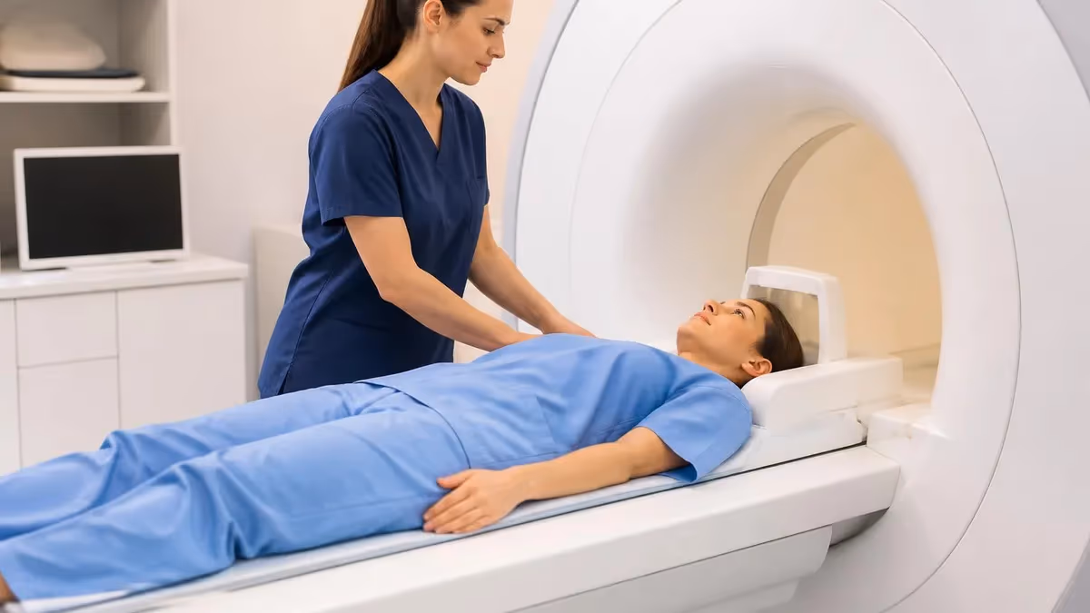
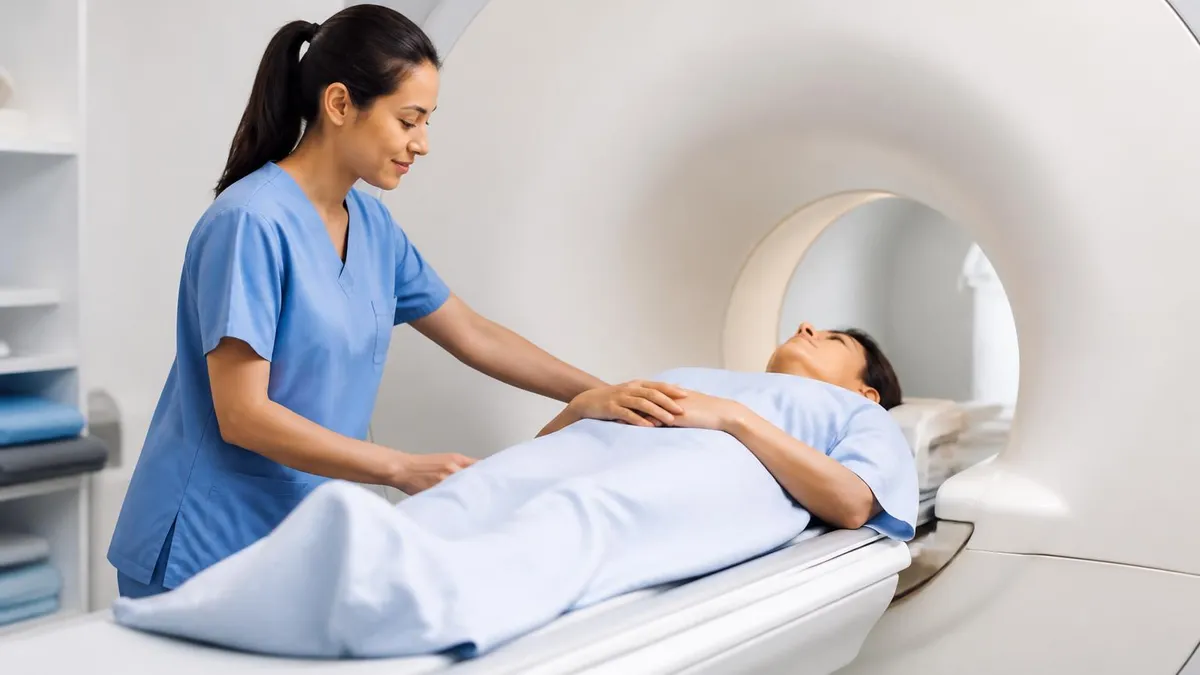
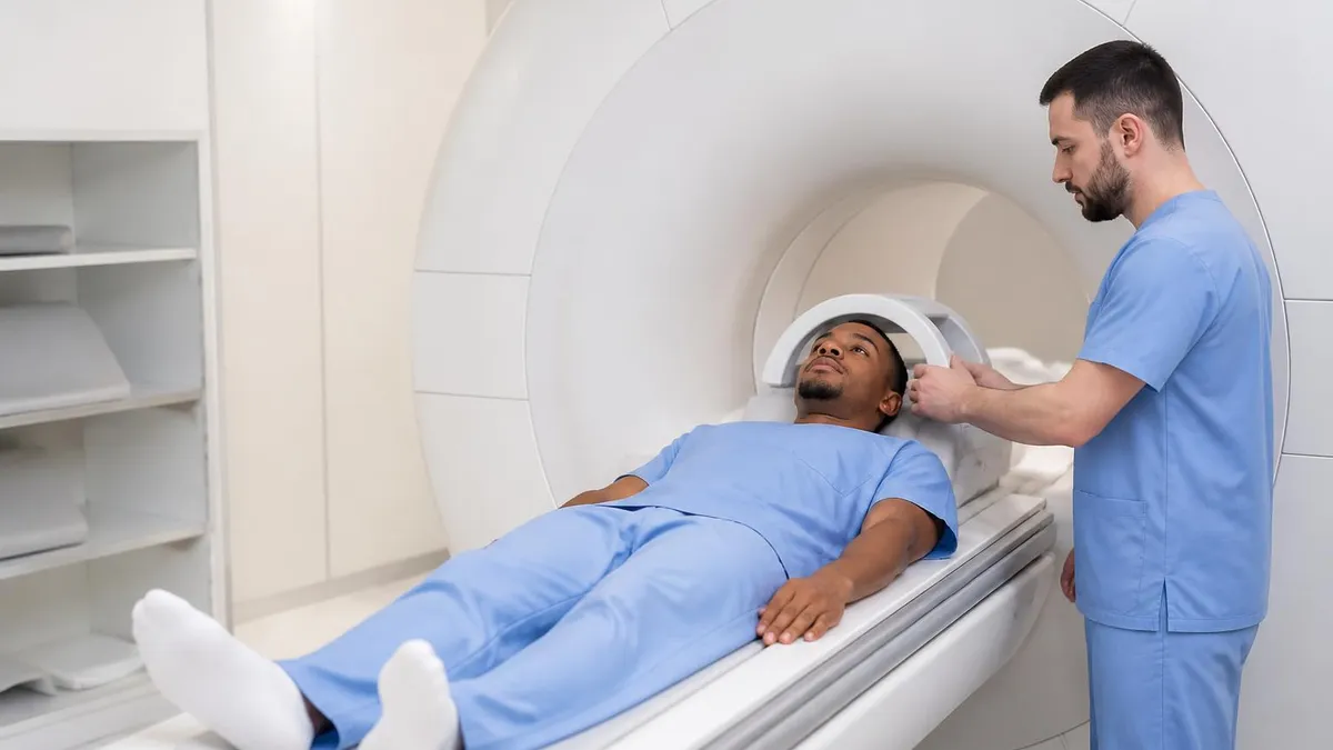
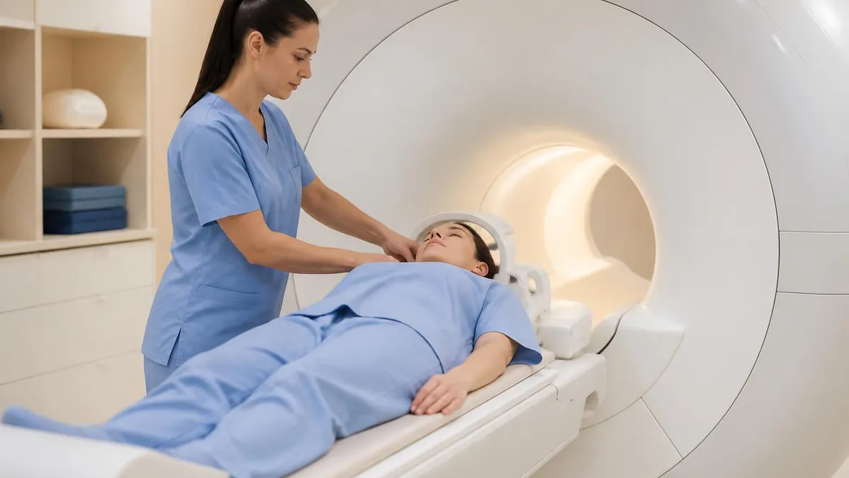
MRI MRA Patient Preparation Checklist
- ✓Confirm no implanted cardiac devices, neurostimulators, or cochlear implants that are MR-unsafe
- ✓Remove all metallic jewelry, hairpins, dentures, and clothing with metal fasteners before entering the scan room
- ✓Disclose any prior surgery involving aneurysm clips, stents, or orthopedic hardware with model numbers
- ✓Fast for four hours before contrast-enhanced studies to reduce risk of nausea after gadolinium injection
- ✓Provide a recent estimated glomerular filtration rate if gadolinium contrast is planned
- ✓Inform the technologist of pregnancy, breastfeeding, or possible pregnancy before scanning begins
- ✓Discuss claustrophobia with the ordering physician and consider mild oral anxiolytic if needed
- ✓Wear loose comfortable clothing or change into the gown the imaging center provides
- ✓Continue prescribed medications with small sips of water unless told otherwise
- ✓Arrive at least fifteen minutes early to complete the MRI safety screening questionnaire
MRA finds most clinically significant disease, but small aneurysms can still hide
Modern 3T MRA detects roughly 95 percent of intracranial aneurysms larger than three millimeters but only about 65 to 75 percent of those under three millimeters. When clinical suspicion remains high after a negative MRA, catheter digital subtraction angiography is still the diagnostic gold standard and should be considered.
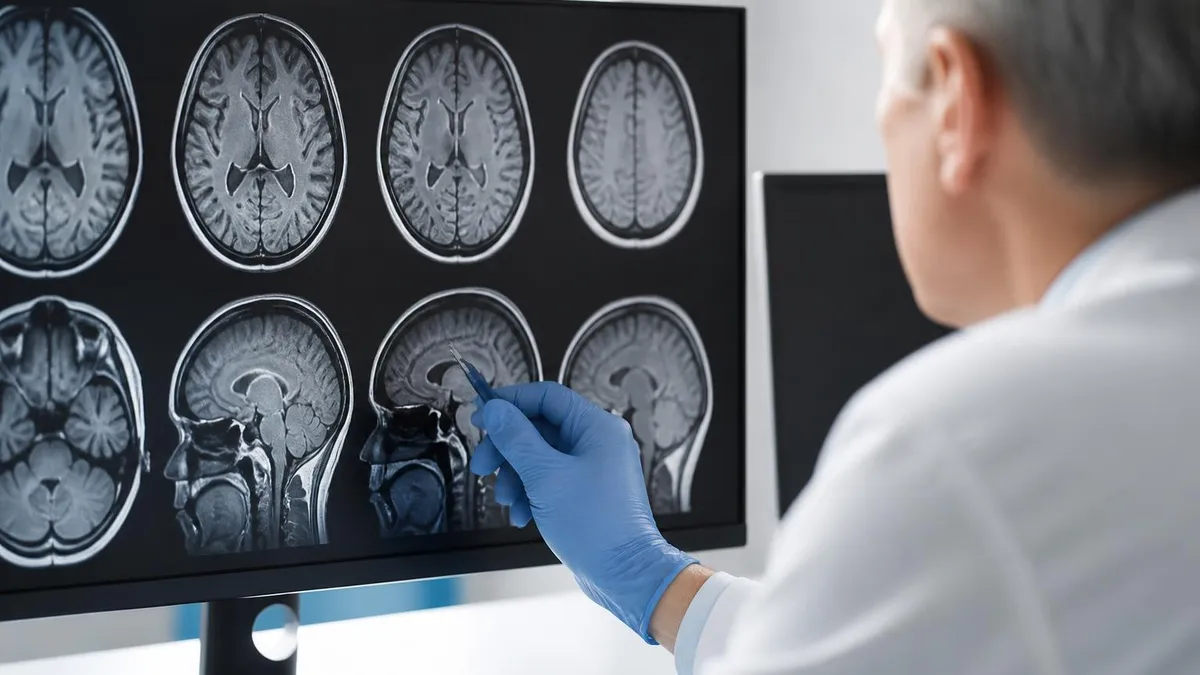
Reading an MRI MRA study begins with the source images, the raw axial slices that contain all of the diagnostic information. Although the colorful maximum-intensity-projection reconstructions are visually striking and useful for presentation, the radiologist relies on the source images to distinguish true narrowing from flow artifacts, to identify intramural hematoma in dissection, and to confirm that signal loss represents pathology rather than turbulent or in-plane flow within a normal vessel that simply does not enhance well.
Reports are structured by vascular territory and typically describe vessel patency, caliber, contour, and any focal lesions. For carotid disease the radiologist states percent stenosis using a recognized method, characterizes plaque morphology when visible, and comments on the contralateral vessel for symmetry. Intracranial reports cover the circle of Willis segment by segment, noting aneurysms with their location, size, neck width, and orientation, all features that influence whether endovascular coiling or surgical clipping is favored by the treating neurosurgeon.
Aortic MRA reports include aortic root diameter, ascending and descending diameters at standardized levels, and the presence of dissection flaps, intramural hematoma, or penetrating ulcer. For surveillance of connective tissue disorders, the report compares current measurements to prior studies and flags any change exceeding two to three millimeters per year. These growth thresholds drive decisions about prophylactic surgical replacement, so consistency between exams is critical and most centers use identical sequences and measurement landmarks each time.
Renal MRA describes the number of renal arteries on each side, the presence of accessory vessels, and any stenosis at the ostium or main trunk. Fibromuscular dysplasia produces a characteristic beaded appearance in the mid to distal main renal artery, while atherosclerotic stenosis tends to involve the ostium. Reports also note kidney size and parenchymal signal because asymmetric atrophy supports clinically significant hemodynamic effect from a stenosis that might otherwise appear borderline on imaging alone.
Peripheral runoff studies are read station by station, from the infrarenal aorta through the iliacs, common femoral, superficial femoral, popliteal, and tibial arteries down to the pedal arch. Each segment is graded as normal, less than fifty percent stenotic, fifty to ninety-nine percent stenotic, or occluded. The radiologist identifies any reconstitution by collaterals, which helps the vascular surgeon plan revascularization targets and decide between balloon angioplasty, stent placement, or surgical bypass.
Artifacts that mimic disease include pulsation ghosting along the phase-encode direction, susceptibility from adjacent bowel gas or surgical clips, and motion from breathing or swallowing during long acquisitions. Experienced readers learn to recognize these patterns rather than calling them pathology. When in doubt, they recommend a confirmatory study such as duplex ultrasound for the carotids or CT angiography for the aorta, especially when the imaging finding would dramatically change patient management or trigger an invasive procedure.
Modern radiology workstations include AI-assisted tools that auto-segment vessels, generate centerlines, and compute stenosis percentages automatically. These tools accelerate reporting but require human verification because they still misregister in cases of dissection, complex aneurysm geometry, and heavy calcification. The final report remains a synthesis of imaging abnormal mri brain context, and prior studies, written by a radiologist who takes responsibility for the interpretation that guides patient care.

Patients with an estimated glomerular filtration rate below 30 milliliters per minute face increased risk of nephrogenic systemic fibrosis from older linear gadolinium agents. Most U.S. centers now use macrocyclic agents, but always verify renal function and discuss risks before any contrast-enhanced MRA in advanced kidney disease.
Choosing between MRA, CT angiography, and duplex ultrasound is one of the most common decisions in vascular imaging. Each modality has strengths that fit particular clinical scenarios, and reasonable clinicians often disagree about the best first test. Understanding the trade-offs helps both ordering providers and patients participate in an informed conversation about which scan to schedule, especially when symptoms are subacute and there is time to optimize the diagnostic pathway rather than acting urgently.
CT angiography is faster, more widely available, and offers higher spatial resolution than MRA. A modern multidetector scanner can image the entire aorta and runoff vessels in under thirty seconds with submillimeter resolution. The trade-offs are ionizing radiation exposure, iodinated contrast with its associated nephrotoxicity and allergy risks, and difficulty imaging vessels obscured by dense calcification, which appears as blooming artifact on CT but is often invisible on MRA. For more details on how MRI compares to other modalities, see our deep dive on What Is an MRI Test? How Magnetic Resonance Imaging Scans Diagnose Disease in 2026.
Duplex ultrasound combines grayscale imaging with Doppler velocity measurement to assess vessels at the bedside. It is portable, inexpensive, free of ionizing radiation, and requires no intravenous contrast. The disadvantages are operator dependence, limited depth penetration in obese patients, and inability to evaluate vessels obscured by bone or bowel gas. Ultrasound shines for carotid screening, lower-extremity deep venous thrombosis, and rapid postoperative checks of bypass grafts and arteriovenous fistulas in dialysis patients across outpatient settings.
For acute stroke workup, most U.S. comprehensive stroke centers use CT angiography because it is fast enough to be completed within the door-to-needle and door-to-puncture metrics that drive treatment outcomes. MRA is more commonly used for subacute evaluation, transient ischemic attack workup in the outpatient setting, and follow-up of known vascular abnormalities where speed is less critical than avoiding repeated radiation exposure over years of surveillance imaging in younger patients with chronic vascular conditions.
For aortic disease in younger patients, especially those with connective tissue disorders, MRA is generally preferred for longitudinal surveillance because these patients require imaging every six to twelve months over decades. Cumulative radiation dose from CT becomes a meaningful concern, and the slightly lower spatial resolution of MRA is offset by reproducibility advantages of using the same modality at every visit. Most thoracic aortic clinics standardize on MRA for this reason and only obtain CT when MRI is contraindicated or unavailable.
For peripheral arterial disease, the choice often depends on local expertise and patient factors. Patients with severe renal impairment, pacemakers, or claustrophobia frequently route to CT angiography or non-contrast MRA techniques. Diabetic patients with heavily calcified tibial vessels may be better served by MRA because calcification does not obscure the lumen as it does on CT, and the resulting images guide endovascular planning more reliably than blooming-affected CT studies in many community practices.
Ultimately, the best vascular imaging strategy is one that combines clinical judgment with knowledge of each modality. MRI MRA earns its place in the diagnostic toolkit because it offers radiation-free, contrast-flexible, multiplanar imaging with quantitative flow capability and excellent tissue characterization. It is not the right test for every patient, but when it is the right test, few alternatives match its combination of safety, diagnostic accuracy, and ability to address both vascular and parenchymal questions at once.
Practical preparation for an MRI MRA exam starts days before the appointment. Patients should gather a list of prior surgeries, implanted devices, and previous imaging studies, and bring this information to the safety screening. If any device card is available, photographing both sides of it and emailing the picture to the imaging center in advance helps the MR safety officer verify compatibility before the patient arrives. This small step can prevent same-day cancellations and the frustration of rescheduling a study that took weeks to coordinate.
On the day of the scan, plan to arrive at least fifteen minutes early. Wear comfortable, loose-fitting clothing without metallic zippers, snaps, or underwire. Many centers provide gowns to ensure no hidden metal interferes with image quality. Leave watches, jewelry, hearing aids, and credit cards in a locker, as the strong static magnetic field can damage magnetic stripes and pull ferromagnetic objects toward the bore with dangerous force. For more on what to expect inside the scan room, see our guide to Noise of MRI Machine: Why MRI Scanners Are So Loud and What to Expect.
Hydration matters for contrast-enhanced studies. Drinking water in the hours before and after gadolinium injection supports renal clearance and may reduce the risk of headache or nausea. Patients should not stop prescribed medications without first consulting the ordering physician, but it is reasonable to take regular pills with a small sip of water on the morning of the exam. Diabetic patients on metformin generally do not need to hold the medication for gadolinium, although institutional protocols vary and should be confirmed in advance.
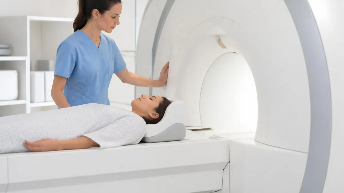
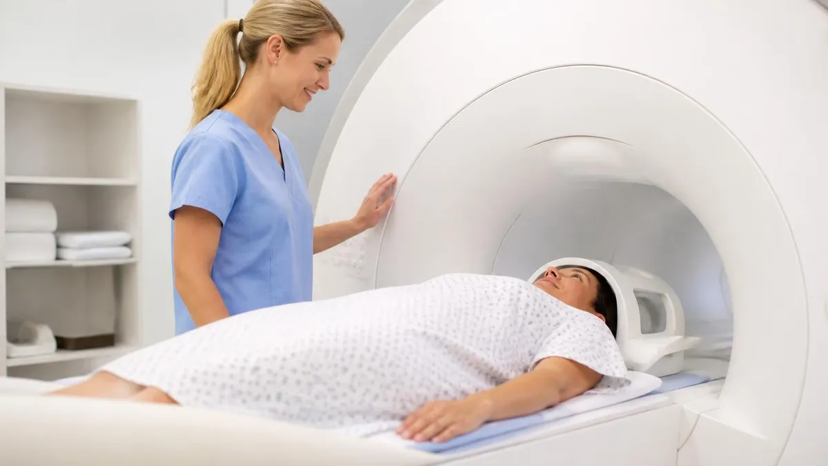
During the scan itself, holding still is the most important contribution a patient can make to image quality. Even small movements during the acquisition of a single angiographic phase can blur the entire vessel tree and require repeating the sequence. Closing the eyes, listening to the offered music, and focusing on slow rhythmic breathing all help. If a breath hold is requested, practice it before the scan begins so the technologist knows how long the patient can comfortably hold and can plan acquisitions around that limit.
Claustrophobia affects roughly five to ten percent of patients undergoing MRI. Options include the use of a mild oral anxiolytic prescribed by the referring physician, scheduling the exam at a center with a wide-bore or open scanner, and using prism glasses that allow the patient to see out of the bore. Telling the technologist about anxiety ahead of time is far better than struggling in silence during the scan. Most centers can pause acquisition, slide the patient partway out, and resume once composure returns.
After the exam, most patients can resume normal activities immediately. There is no recovery period for MRA. If gadolinium was administered, hydration through the rest of the day supports clearance. Breastfeeding patients may continue without interruption under current 2026 guidelines from major U.S. radiology societies, which consider gadolinium transfer into breast milk negligible. The radiologist typically issues a report within twenty-four to forty-eight hours, and the ordering physician will discuss findings at the next clinic visit or sooner if urgent.
Understanding the test, preparing thoughtfully, and communicating openly with the imaging team turn a potentially intimidating exam into a routine diagnostic step. MRI MRA delivers detailed vascular information without radiation, without arterial puncture, and often without injected contrast, and a well-prepared patient gets the most diagnostic value out of every minute spent inside the scanner during the appointment.
MRI Questions and Answers

What Is an MRI Test? How Magnetic Resonance Imaging Scans Diagnose Disease in 2026
MRI Medical Abbreviation: What MRI Stands For and Why It Matters
The History of MRI: From Discovery to Modern Medicine
Knee MRI Images: A Complete Guide to Reading, Understanding, and Interpreting Knee Scans
Noise of MRI Machine: Why MRI Scanners Are So Loud and What to Expect
About the Author
Medical Laboratory Scientist & Clinical Certification Expert
Johns Hopkins UniversityDr. Sandra Kim holds a PhD in Clinical Laboratory Science from Johns Hopkins University and is certified as a Medical Technologist (MT) and Medical Laboratory Scientist (MLS) through ASCP. With 16 years of clinical laboratory experience spanning hematology, microbiology, and molecular diagnostics, she prepares candidates for ASCP board exams, MLT, MLS, and specialist certification tests.
Join the Discussion
Connect with other students preparing for this exam. Share tips, ask questions, and get advice from people who have been there.
View discussion (6 replies)