Anatomy and Physiology Overview: Body Systems, Core Concepts, and Professional Practice
🧠 Free Anatomy and Physiology Overview: Body practice test with questions and answer explanations. Prepare for the 2026 July exam with instant scoring.
Anatomy and physiology are the two foundational sciences that underpin all clinical healthcare practice. Anatomy describes structure — where body parts are located, how they're organized, and how they relate to each other spatially. Physiology describes function — how those structures work, how they maintain homeostasis, and how they respond to stress, injury, and intervention. Understanding both together is what allows a clinician, therapist, or trainer to interpret symptoms, design safe interventions, and communicate effectively with other healthcare providers.
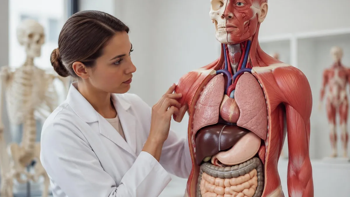
The subject spans every level of biological organization: from chemical bonds and cellular membranes up through tissues, organs, and organ systems, all the way to the whole organism interacting with its environment. In professional practice contexts — whether massage therapy, physical therapy, personal training, nursing, or any allied health field — anatomy and physiology knowledge isn't abstract. It's what tells you why a muscle might be hypertonic, why a joint loses range of motion after immobilization, or why a client's cardiovascular response to exercise changes with age.
This overview covers the structural and functional principles that appear most frequently in professional certification exams and clinical practice: body organization and anatomical terminology, the musculoskeletal system in depth, core physiological concepts including homeostasis and feedback mechanisms, applied assessment techniques, and professional standards for communication, documentation, and continuing education. Whether you're preparing for a certification exam or deepening clinical knowledge, these are the domains that matter most in applied anatomy and physiology practice.
Structural organization in anatomy follows a hierarchical sequence: chemical level (atoms and molecules, including water, proteins, lipids, and nucleic acids), cellular level (the basic functional unit of life), tissue level (four primary types: epithelial, connective, muscle, and nervous), organ level (structures with two or more tissue types performing a specific function), organ system level (multiple organs with related functions), and organismal level (all systems working together). Every clinical observation locates something on this hierarchy — understanding where a pathology or dysfunction sits determines the appropriate intervention.
Anatomical terminology uses standardized directional and positional language to describe location precisely. Medial means toward the midline; lateral means away from it. Superior is toward the head; inferior is toward the feet. Anterior (ventral) means toward the front; posterior (dorsal) means toward the back. Proximal is closer to the point of attachment or origin; distal is further away. Superficial refers to structures closer to the body surface; deep refers to structures further from it. These terms are always described relative to anatomical position — the body standing upright, arms at sides, palms facing forward.
Body planes divide the body for anatomical description and imaging reference. The sagittal plane divides the body into left and right sections — a midsagittal (median) plane divides it equally. The frontal (coronal) plane divides the body into anterior and posterior sections. The transverse (horizontal) plane divides the body into superior and inferior sections. CT scans are typically displayed in transverse cross-sections; MRI can be reformatted in any plane. Understanding planes is essential for interpreting imaging findings and for describing joint motion in three-dimensional space.
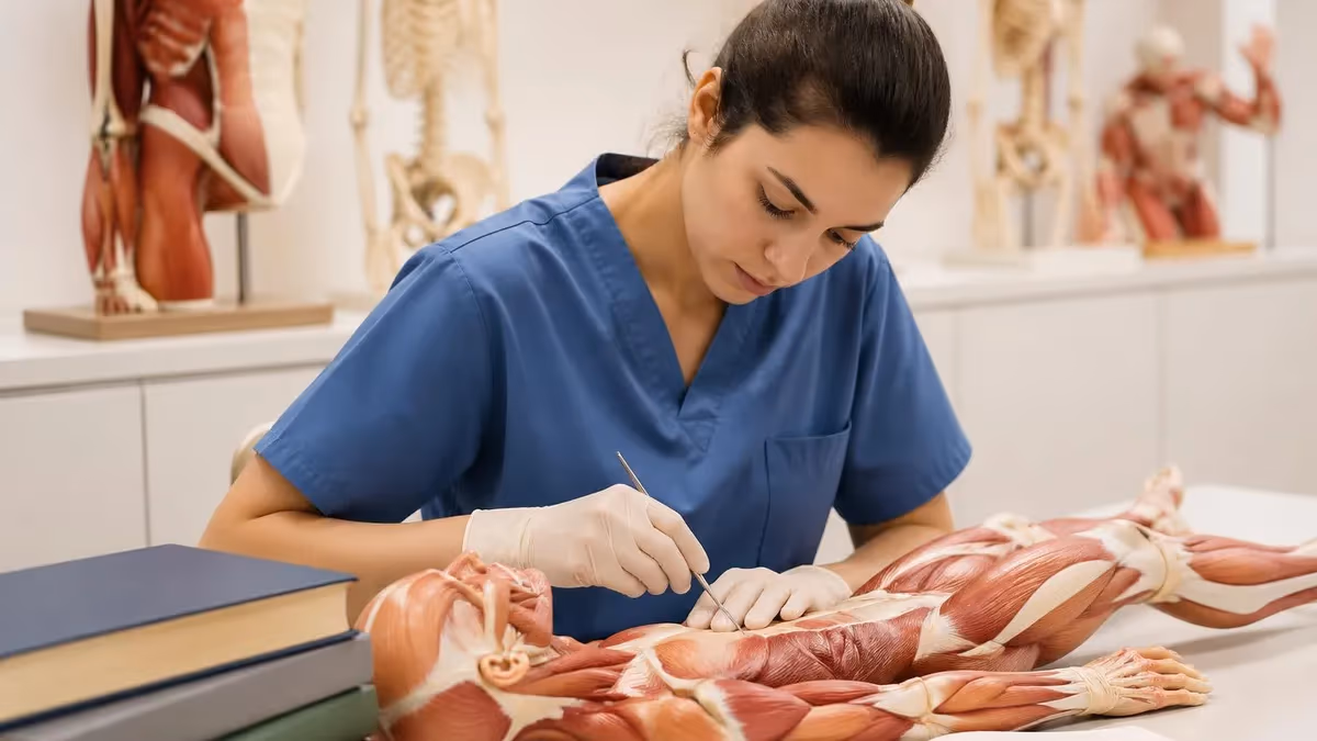
The musculoskeletal system is the largest and most clinically relevant body system for movement-based professionals. It consists of bones, cartilage, joints, ligaments, tendons, and skeletal muscle — working together to produce movement, support the body against gravity, protect internal organs, and store and release metabolic energy. The skeleton provides rigid levers; muscles generate force through contraction; joints define the axes and ranges of motion; connective tissues transmit and modulate forces throughout the system.
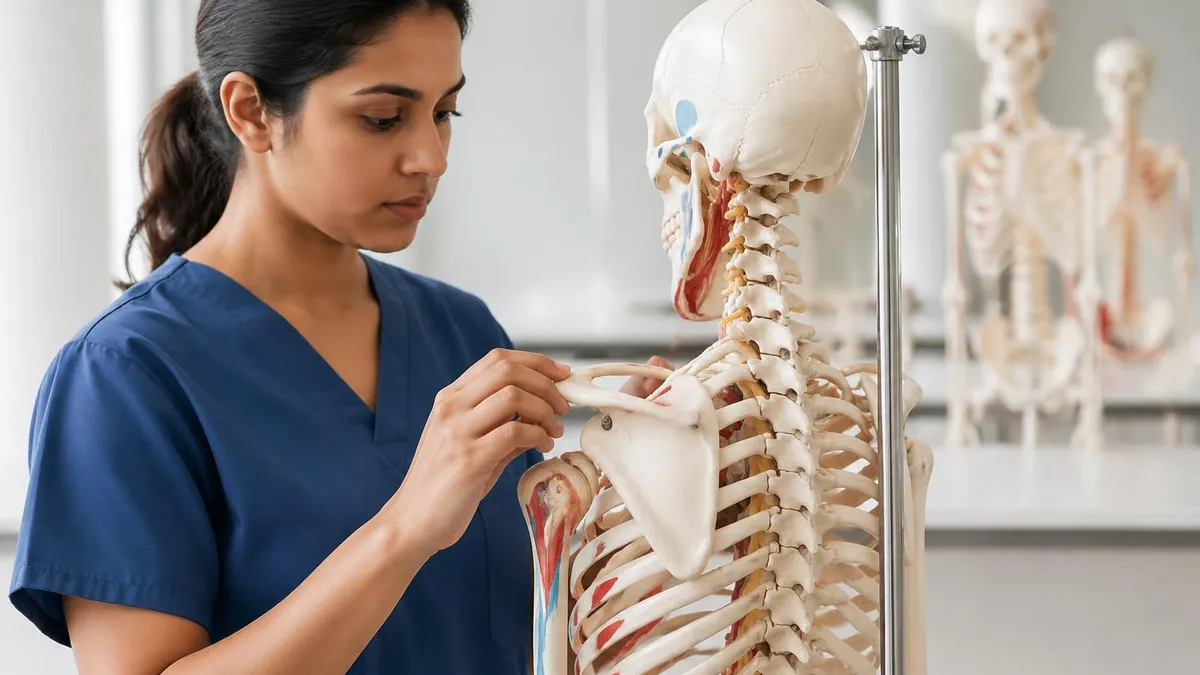
Bone tissue is metabolically active throughout life, constantly remodeling in response to mechanical loading, hormonal signaling, and nutritional status. Cortical (compact) bone forms the dense outer shell of long bones, providing resistance to bending. Cancellous (trabecular) bone fills the interior with a porous network aligned along stress lines, balancing strength with lightness.
Joints are classified by their structural composition (fibrous, cartilaginous, or synovial) and by their functional range of motion. Synovial joints — the freely movable diarthroses — are the most clinically relevant: they have a joint capsule filled with synovial fluid, articular cartilage covering the opposing bone surfaces, and a spectrum of stabilizing ligaments specific to each joint.
Skeletal muscle is organized hierarchically: the whole muscle (enclosed in epimysium) contains fascicles (enclosed in perimysium), which contain individual muscle fibers (enclosed in endomysium), which contain myofibrils made of sarcomeres. Contraction occurs at the sarcomere level when actin and myosin filaments slide past each other — the sliding filament theory. Muscle force production depends on the number of motor units recruited, the firing frequency of those units, muscle length at the time of contraction, and the velocity of contraction. Eccentric contractions (muscle lengthening under load) produce the most force and the most delayed onset muscle soreness (DOMS).
Major Body Systems Overview
The nervous system integrates sensory input, processes information, and coordinates motor output. The central nervous system (brain and spinal cord) processes and integrates; the peripheral nervous system (cranial and spinal nerves) transmits signals to and from the CNS. The autonomic nervous system — divided into sympathetic (fight-or-flight) and parasympathetic (rest-and-digest) branches — regulates involuntary functions including heart rate, digestion, and glandular secretion. Clinical relevance: nerve compression (radiculopathy), autonomic dysregulation, and dermatome mapping for locating injury level.
The endocrine system communicates through hormones — chemical messengers released into the bloodstream to reach distant target cells. Major glands include the pituitary (master gland, controls other endocrine glands), thyroid (metabolic rate), adrenal cortex (cortisol, stress response; aldosterone, fluid balance), pancreas (insulin and glucagon for blood glucose regulation), and gonads (sex hormones). Endocrine-musculoskeletal interactions are clinically significant: low estrogen accelerates bone loss, chronic cortisol elevation impairs tissue repair, and growth hormone drives musculoskeletal development.
Homeostasis — the maintenance of a relatively stable internal environment despite changing external conditions — is the central organizing principle of physiology. The body maintains homeostasis through feedback mechanisms: negative feedback loops dominate (the correction opposes the deviation, returning the variable toward its set point), while positive feedback loops amplify deviations toward a physiological goal (childbirth contractions, blood clotting, nerve action potentials). Understanding feedback mechanisms is essential for interpreting how the body responds to exercise, injury, and therapeutic intervention.
The cell is the fundamental unit of life, and cellular physiology underpins every system-level process. Key cellular functions include metabolism (catabolism breaks molecules down for energy; anabolism builds molecules from smaller components), membrane transport (passive diffusion, facilitated diffusion, active transport, osmosis), communication (receptors for hormones, neurotransmitters, and growth factors), and the cell cycle (growth, DNA replication, division). In clinical contexts, cellular physiology explains why tissue healing follows predictable phases, why overuse injuries occur at specific anatomical sites, and why certain interventions accelerate or impair recovery.
Tissue types determine how structures respond to mechanical stress, injury, and intervention. Epithelial tissue covers surfaces and lines cavities — it heals quickly because cells divide rapidly. Connective tissue (including bone, cartilage, tendons, ligaments, and adipose) provides structural support — it heals more slowly due to lower vascularity. Muscle tissue generates force through contraction — skeletal muscle is voluntary and can regenerate to a limited degree; cardiac muscle is involuntary and regenerates poorly after injury. Nervous tissue transmits electrical impulses — peripheral nerves can regenerate slowly (1-3 mm/day) if the cell body is intact; CNS neurons do not regenerate effectively.
Applied anatomy methods translate structural knowledge into clinical assessment and intervention. Palpation — systematic tactile examination — requires knowing precise anatomical landmarks: bony prominences (spinous processes, greater trochanter, ASIS, lateral epicondyle), muscle bellies and their attachments, tendons, ligament positions, and nerve trunk locations. Effective palpation requires relaxed tissue (patient positioned to minimize muscle tension), correct depth of pressure for the target structure, and the ability to distinguish normal tissue texture from hypertonicity, fibrosis, or edema.
Range of motion (ROM) assessment applies anatomical knowledge of joint structure to clinical measurement. Goniometry uses specific anatomical landmarks as reference points — for shoulder abduction, the fulcrum sits at the acromion process, the stationary arm aligns with the lateral midline of the thorax, and the moving arm aligns with the lateral midline of the humerus toward the lateral epicondyle. Deviation from normative values (shoulder abduction 180°, hip flexion 120°, ankle dorsiflexion 20°) points toward specific structural limitations including capsular patterns, muscle shortening, or joint pathology.
Manual muscle testing (MMT) assesses the strength and function of specific muscles by applying resistance in the direction opposite to the muscle's action, with the patient positioned to isolate the target muscle. Grading uses a 0-5 scale: 0 = no contraction, 1 = visible contraction without movement, 2 = full ROM gravity-eliminated, 3 = full ROM against gravity, 4 = full ROM against moderate resistance, 5 = full ROM against maximum resistance. MMT requires knowledge of each muscle's origin, insertion, action, and the correct positioning to isolate it from synergists.
Textbook vs. Visual/3D Anatomy Learning
- +Anatomy textbooks provide comprehensive, systematically organized reference content
- +3D software (Complete Anatomy, Visible Body) allows rotation and layer-by-layer dissection
- +Cadaveric study reveals variation and spatial relationships that images can't fully convey
- +Flashcard systems (Anki) build retrieval-based learning that improves exam performance
- +Clinical case studies integrate anatomy knowledge with real patient presentations
- +Video dissection series (Acland's, Getty Images Medical) are more accessible than cadaver lab access
- −Textbooks present idealized anatomy that varies significantly in real patients
- −3D software can give false confidence without hands-on palpation practice
- −Cadaver lab access is restricted to formal educational programs in most jurisdictions
- −Isolated memorization without clinical application produces knowledge that doesn't transfer
- −Over-reliance on 2D images doesn't build the three-dimensional spatial reasoning clinical work requires
- −Without spaced repetition, anatomy knowledge decays rapidly without active clinical reinforcement
Clinical communication in anatomy-based professions requires precise use of anatomical terminology — not because formal language is required for its own sake, but because ambiguous descriptions of structure and location create real clinical risk. Saying "the patient's shoulder hurts" tells the next provider almost nothing clinically relevant.
Saying "the patient reports pain at the anterior aspect of the glenohumeral joint with active elevation above 90 degrees, reproduced on palpation of the long head biceps tendon at the intertubercular groove" communicates location, behavior, and probable structure — and it allows the receiving provider to build on that assessment rather than starting from scratch.
Documentation follows the same principle. SOAP notes (Subjective, Objective, Assessment, Plan) organize clinical findings into a structured format: what the patient reports (S), what you observe and measure (O), what those findings indicate (A), and what you're going to do about it (P). In the Objective section, anatomical precision is essential — ROM measurements with specific joints and planes, palpation findings with named structures, postural observations with anatomical landmarks. Vague documentation creates medicolegal exposure and impedes continuity of care when other providers see the patient.
Interprofessional communication relies on shared anatomical vocabulary. When a massage therapist refers to a physician, the referral letter's clinical value depends on whether it communicates findings in terms the physician can interpret. Conversely, a physician's referral to a physical therapist or massage therapist is more useful if it specifies anatomical diagnosis rather than just symptoms. Building fluency in standard anatomical and clinical terminology is one of the highest-yield professional development activities for any hands-on healthcare provider.
10 Anatomy and Physiology Study Strategies
- ✓Use spaced repetition (Anki flashcards) for anatomical terminology — retrieval practice beats re-reading
- ✓Study each muscle with all five attributes: origin, insertion, action, innervation, and blood supply
- ✓Practice palpating anatomical landmarks on a partner or model, not just on diagrams
- ✓Connect structure to function — always ask why a structure is shaped the way it is
- ✓Draw the musculoskeletal system from memory repeatedly — active recall builds retention better than passive review
- ✓Use 3D anatomy software to visualize structures from multiple planes and layers
- ✓Practice goniometric measurement on willing partners to build muscle memory for correct landmark placement
- ✓Study pathology alongside normal anatomy — understanding dysfunction deepens understanding of normal function
- ✓Review dermatomes and myotomes separately — they appear on exams and are clinically essential for neurological assessment
- ✓Connect your studying to clinical scenarios: how would you use this knowledge with a real patient?
The musculoskeletal system's functional anatomy extends beyond individual muscles to movement patterns. Synergists assist the prime mover (agonist) in producing a movement. Antagonists oppose the prime mover and must relax for movement to occur efficiently — chronic antagonist tightness is a common source of movement dysfunction. Stabilizers co-contract to maintain joint position while the prime movers act on adjacent joints. In the shoulder, the rotator cuff muscles (supraspinatus, infraspinatus, teres minor, subscapularis) function primarily as dynamic stabilizers of the glenohumeral joint, preventing superior translation of the humeral head during arm elevation — even though they also produce rotation.
Kinetic chain concepts describe how forces are transmitted through interconnected segments. A closed kinetic chain exercise has the distal segment fixed (foot on ground during a squat); an open kinetic chain exercise has the distal segment free (leg extension machine). Closed kinetic chain exercises are generally more functional and produce less shear force on joints like the ACL. Understanding kinetic chain function explains why a hip weakness can manifest as knee pain, or why limited ankle dorsiflexion drives compensatory patterns up through the lumbar spine.
Connective tissue mechanics are particularly important for manual therapists. Tendons transmit muscle force to bone — they're highly tensile but poorly vascularized, which is why tendinopathies heal slowly. Ligaments stabilize joints by limiting excessive motion — sprains damage ligament fibers in grades: Grade I (microscopic tearing, intact ligament), Grade II (partial tear), Grade III (complete rupture). Fascia is a continuous sheet of connective tissue that envelops and compartmentalizes every structure in the body — fascial restrictions can transmit tension across anatomical regions and are the rationale for myofascial release techniques.
Continuing education in anatomy-based professions isn't just a licensing requirement — it's a clinical necessity, because the research base changes meaningfully over five-to-ten-year periods. The understanding of fascia has been transformed by research conducted after 2000. Pain neuroscience education has shifted the clinical framing of chronic pain from structural to neurophysiological. Updated biomechanical models have changed recommendations for exercise technique and therapeutic loading. Providers who stop updating their anatomy and physiology knowledge after initial certification are working from an increasingly outdated clinical model.
Industry best practices in anatomy-based professions share several common threads: evidence-informed assessment before intervention, clear goal-setting based on objective findings, regular reassessment to evaluate progress, appropriate referral when findings exceed scope of practice, and accurate documentation. The best practice standard isn't perfection — it's systematic application of current evidence to clinical decision-making. Professional associations publish practice guidelines and continuing education curricula that operationalize these standards in profession-specific language.
Scope of practice is a critical professional boundary that anatomy knowledge helps define. Understanding when a clinical finding falls within your scope (a hypertonic muscle appropriate for manual therapy) versus outside it (a tumor mass requiring medical evaluation) requires both anatomical knowledge and professional judgment. Differential diagnosis — systematically considering multiple explanations for a clinical finding — is the process that maps anatomical knowledge onto scope decisions. Anatomy education that includes pathology recognition alongside normal function prepares providers to recognize when referral is appropriate.
Postural assessment uses anatomical landmarks as objective reference points for identifying deviations from ideal alignment. From the lateral view, a plumb line through the ear, shoulder (acromion), hip (greater trochanter), knee (slightly anterior to knee center), and ankle (slightly anterior to lateral malleolus) represents neutral alignment. Deviations — forward head position, thoracic kyphosis, lumbar hyperlordosis, anterior pelvic tilt — are described using standard anatomical terms and linked to the muscles and joints that drive them.
Gait analysis applies anatomy to dynamic movement. The gait cycle has stance phase (foot on ground, ~60% of cycle) and swing phase (foot off ground, ~40%). During stance, hip extensors decelerate forward trunk momentum at initial contact; knee extensors control flexion; ankle plantar flexors generate propulsion at push-off. Deviations — Trendelenburg gait (hip abductor weakness), antalgic gait (pain avoidance), steppage gait (ankle dorsiflexion weakness) — map directly onto specific muscle or nerve dysfunction and guide targeted assessment.
Special orthopedic tests extend physical examination by applying specific stresses to anatomical structures to elicit or reproduce symptoms. Each test has a target tissue (the structure being stressed), a provocative mechanism (the direction and type of force applied), and a positive finding (pain, instability, or reproduction of symptoms). Sensitivity (ability to detect true positives) and specificity (ability to rule out true negatives) vary widely between tests. No single special test is diagnostic in isolation — findings must be interpreted within the full clinical picture including history, postural assessment, ROM, and strength testing.
Anatomy Practice Test Questions
Prepare for the Anatomy Exam exam with our free practice test modules. Each quiz covers key topics to help you pass on your first try.
Anatomy of the Heart
Anatomy Exam Questions covering Anatomy of the Heart. Master Anatomy Test concepts for certification prep.
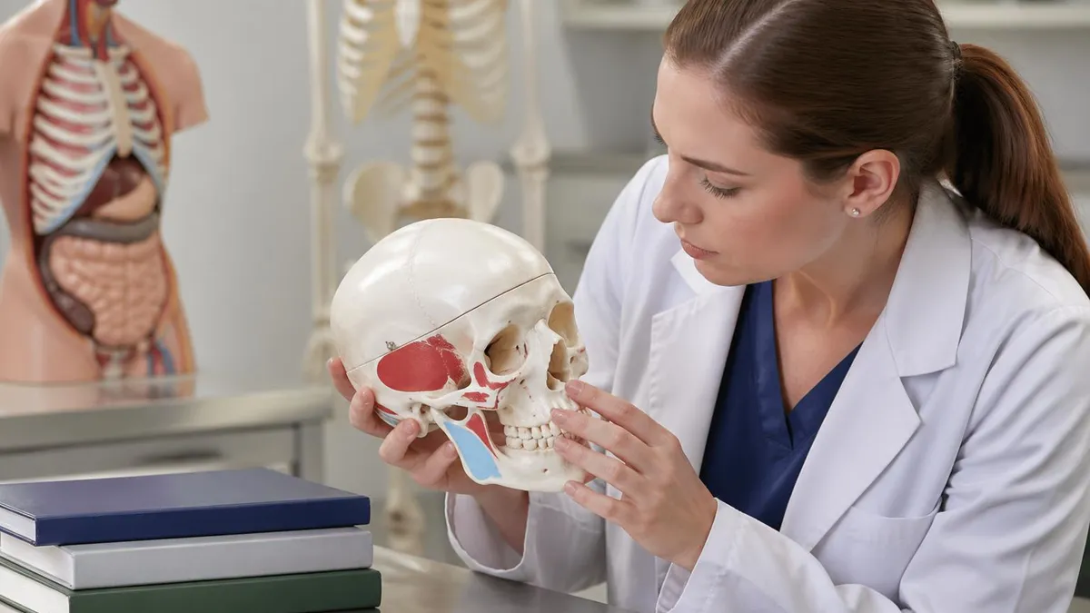
Anatomy Bones of the Cranial Vault
Free Anatomy Practice Test featuring Bones of the Cranial Vault. Improve your Anatomy Exam score with mock test prep.
Anatomy Brachial Plexus Structures
Anatomy Mock Exam on Brachial Plexus Structures. Anatomy Study Guide questions to pass on your first try.
Anatomy Cranial Nerves and Foramina
Anatomy Test Prep for Cranial Nerves and Foramina. Practice Anatomy Quiz questions and boost your score.
Anatomy Exam Anatomy Lymphatic System
Anatomy Questions and Answers on Exam Anatomy Lymphatic System. Free Anatomy practice for exam readiness.
Anatomy Exam Anatomy Muscles of the Lower ...
Anatomy Mock Test covering Exam Anatomy Muscles of the Lower Limb. Online Anatomy Test practice with instant feedback.
Anatomy Gross Anatomy of the Kidney
Free Anatomy Quiz on Gross Anatomy of the Kidney. Anatomy Exam prep questions with detailed explanations.
Anatomy Histology of Connective Tissues
Anatomy Practice Questions for Histology of Connective Tissues. Build confidence for your Anatomy certification exam.
Anatomy Joints of the Lower Limb
Anatomy Test Online for Joints of the Lower Limb. Free practice with instant results and feedback.
Anatomy Muscles of the Rotator Cuff
Anatomy Study Material on Muscles of the Rotator Cuff. Prepare effectively with real exam-style questions.
Anatomy Upper Gastrointestinal Tract
Free Anatomy Test covering Upper Gastrointestinal Tract. Practice and track your Anatomy exam readiness.
Anatomy and Physiology
Anatomy Exam Questions covering and Physiology. Master Anatomy Test concepts for certification prep.
Anatomy and physiology knowledge doesn't become clinically useful until you can apply it under time pressure with a real patient in front of you. The gap between knowing anatomy in isolation and deploying it during assessment is real — and it closes through practice, not through more reading.
The most effective approach is integrating anatomy study with clinical skill practice: learn a muscle group, immediately practice palpating it on a partner, then design and practice an assessment for common dysfunctions in that region. That integration loop accelerates both retention and clinical application far more effectively than studying anatomy and practicing skills as separate activities.
For certification exam preparation specifically, prioritize the musculoskeletal system (most questions in movement-based certification exams), the nervous system (dermatomes, myotomes, and autonomic function), and the cardiovascular and respiratory systems (for contraindication and precaution knowledge). Then layer in the professional practice content — communication, documentation, scope of practice, and continuing education — which requires less anatomical depth but more understanding of professional context and standards.
The deeper value of anatomy and physiology mastery is that it builds clinical confidence. Providers who understand the structural and functional basis for their interventions can explain them to clients clearly, modify them intelligently when standard approaches don't work, recognize when something unexpected in a session suggests referral, and continue developing their practice as the research base evolves. Anatomy isn't a subject you finish learning — it's a framework that grows more useful the more clinical experience you bring to it.
Anatomy and Physiology Questions and Answers
About the Author

Educational Psychologist & Academic Test Preparation Expert
Columbia University Teachers CollegeDr. Lisa Patel holds a Doctorate in Education from Columbia University Teachers College and has spent 17 years researching standardized test design and academic assessment. She has developed preparation programs for SAT, ACT, GRE, LSAT, UCAT, and numerous professional licensing exams, helping students of all backgrounds achieve their target scores.